 Movie
Movie Controller

[English] 日本語
 Yorodumi
- PDB-3eo1: Structure of the Fab Fragment of GC-1008 in Complex with Transfor... -
+ Open data

- Basic information
| Entry | Database: PDB / ID: 3eo1 | ||||||
|---|---|---|---|---|---|---|---|
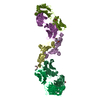
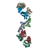
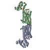
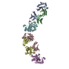
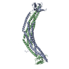
| Title | Structure of the Fab Fragment of GC-1008 in Complex with Transforming Growth Factor-Beta 3 | ||||||
 Components Components |
| ||||||
 Keywords Keywords | IMMUNE SYSTEM/CYTOKINE / Antibody-cytokine complex / Growth factor / FAB fragment / Cardiomyopathy / Cleavage on pair of basic residues / Glycoprotein / Mitogen / Polymorphism / Secreted / IMMUNE SYSTEM-CYTOKINE COMPLEX | ||||||
| Function / homology |  Function and homology information Function and homology informationuterine wall breakdown / detection of hypoxia / type III transforming growth factor beta receptor binding / negative regulation of macrophage cytokine production / secondary palate development / positive regulation of tight junction disassembly / type II transforming growth factor beta receptor binding / cell-cell junction organization / type I transforming growth factor beta receptor binding / mammary gland development ...uterine wall breakdown / detection of hypoxia / type III transforming growth factor beta receptor binding / negative regulation of macrophage cytokine production / secondary palate development / positive regulation of tight junction disassembly / type II transforming growth factor beta receptor binding / cell-cell junction organization / type I transforming growth factor beta receptor binding / mammary gland development / transforming growth factor beta binding / odontogenesis / lung alveolus development / face morphogenesis / positive regulation of filopodium assembly / immunoglobulin complex, circulating / Molecules associated with elastic fibres / Classical antibody-mediated complement activation / immunoglobulin receptor binding / IgG immunoglobulin complex / Initial triggering of complement / TGF-beta receptor signaling activates SMADs / positive regulation of collagen biosynthetic process / positive regulation of cell division / positive regulation of SMAD protein signal transduction / FCGR activation / negative regulation of vascular associated smooth muscle cell proliferation / complement activation, classical pathway / ECM proteoglycans / Role of phospholipids in phagocytosis / positive regulation of epithelial to mesenchymal transition / salivary gland morphogenesis / antigen binding / positive regulation of stress fiber assembly / response to progesterone / transforming growth factor beta receptor signaling pathway / platelet alpha granule lumen / FCGR3A-mediated IL10 synthesis / Regulation of Complement cascade / cytokine activity / B cell receptor signaling pathway / positive regulation of protein secretion / growth factor activity / FCGR3A-mediated phagocytosis / Regulation of actin dynamics for phagocytic cup formation / Platelet degranulation / regulation of cell population proliferation / heart development / antibacterial humoral response / in utero embryonic development / extracellular matrix / Interleukin-4 and Interleukin-13 signaling / blood microparticle / negative regulation of neuron apoptotic process / adaptive immune response / response to hypoxia / positive regulation of apoptotic process / negative regulation of cell population proliferation / positive regulation of cell population proliferation / positive regulation of DNA-templated transcription / positive regulation of transcription by RNA polymerase II / : / extracellular exosome / extracellular region / identical protein binding / plasma membrane Similarity search - Function | ||||||
| Biological species |   Homo sapiens (human) Homo sapiens (human) | ||||||
| Method |  X-RAY DIFFRACTION / SYNCHROTRON / MOLECULAR REPLACEMENT / Resolution: 3.1 Å X-RAY DIFFRACTION / SYNCHROTRON / MOLECULAR REPLACEMENT / Resolution: 3.1 Å | ||||||
 Authors Authors | Gruetter, C. / Gruetter, M.G. | ||||||
 Citation Citation | Journal: Proc.Natl.Acad.Sci.Usa / Year: 2008 Title: A cytokine-neutralizing antibody as a structural mimetic of 2 receptor interactions Authors: Wilkinson, T. / Turner, R. / Podichetty, S. / Finch, D. / McCourt, M. / Loning, S. / Jermutus, L. | ||||||
| History |
|
- Structure visualization
| Structure viewer | Molecule: MolmilJmol/JSmol |
|---|
- Downloads & links
-Download
| PDBx/mmCIF format | 3eo1.cif.gz | 376.9 KB | Display | PDBx/mmCIF format |
|---|---|---|---|---|
| PDB format | pdb3eo1.ent.gz | 315.8 KB | Display | PDB format |
| PDBx/mmJSON format | 3eo1.json.gz | Tree view | PDBx/mmJSON format | |
| Others |  Other downloads Other downloads |
-Validation report
| Arichive directory | https://data.pdbj.org/pub/pdb/validation_reports/eo/3eo1ftp://data.pdbj.org/pub/pdb/validation_reports/eo/3eo1 | HTTPS FTP |
|---|
-Related structure data
| Related structure data |  3eo0SC  1tgjS S: Starting model for refinement C: citing same article ( |
|---|---|
| Similar structure data |


-Links
 PDBj
PDBj


- Assembly
| Deposited unit | 
| ||||||||
|---|---|---|---|---|---|---|---|---|---|
| 1 | 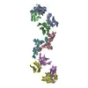
| ||||||||
| 2 | 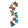
| ||||||||
| Unit cell |
|
-Components
| #1: Antibody | Mass: 23425.934 Da / Num. of mol.: 4 Source method: isolated from a genetically manipulated source Source: (gene. exp.) #2: Antibody | Mass: 23838.779 Da / Num. of mol.: 4 Source method: isolated from a genetically manipulated source Source: (gene. exp.) #3: Protein | Mass: 12734.504 Da / Num. of mol.: 4 / Fragment: UNP residues 301-412, receptor binding fragment Source method: isolated from a genetically manipulated source Source: (gene. exp.) Homo sapiens (human) / Gene: TGFB3 / Production host:  Has protein modification | Y | Sequence details | A SEQUENCE DATABASE REFERENCE FOR THIS PROTEIN (ANTIBODY) DOES NOT CURRENTLY EXIST. | |
|---|
-Experimental details
-Experiment
| Experiment | Method: X-RAY DIFFRACTION / Number of used crystals: 1 |
|---|
- Sample preparation
| Crystal | Density Matthews: 3.47 Å3/Da / Density % sol: 64.5 % |
|---|---|
| Crystal grow | Temperature: 277 K / Method: vapor diffusion, sitting drop / pH: 6.8 Details: 23% tert.-butanol, 0.1M sodium citrate, 0.2M phenoxyacetic acid, pH6.8, VAPOR DIFFUSION, SITTING DROP, temperature 277K |
-Data collection
| Diffraction | Mean temperature: 90 K |
|---|---|
| Diffraction source | Source: SYNCHROTRON / Site: SLS  / Beamline: X06SA / Wavelength: 1.070645 Å / Beamline: X06SA / Wavelength: 1.070645 Å |
| Detector | Type: MARRESEARCH / Detector: CCD / Date: Sep 24, 2006 / Details: BENT MIRRORS |
| Radiation | Monochromator: SI(111) / Protocol: SINGLE WAVELENGTH / Monochromatic (M) / Laue (L): M / Scattering type: x-ray |
| Radiation wavelength | Wavelength: 1.070645 Å / Relative weight: 1 |
| Reflection | Resolution: 3.1→35 Å / Num. obs: 60296 / % possible obs: 97.9 % / Redundancy: 5 % / Rmerge(I) obs: 0.092 / Rsym value: 0.077 / Net I/σ(I): 20.8 |
| Reflection shell | Resolution: 3.1→3.3 Å / Redundancy: 5 % / Rmerge(I) obs: 0.367 / Mean I/σ(I) obs: 3.8 / Num. unique all: 10322 / Rsym value: 0.522 / % possible all: 96.4 |
- Processing
| Software |
| ||||||||||||||||||||
|---|---|---|---|---|---|---|---|---|---|---|---|---|---|---|---|---|---|---|---|---|---|
| Refinement | Method to determine structure: MOLECULAR REPLACEMENT Starting model: PDB ENTRY 3EO0, 1TGJ Resolution: 3.1→35 Å / Stereochemistry target values: MAXIMUM LIKELIHOOD
| ||||||||||||||||||||
| Displacement parameters | Biso mean: 80.7 Å2 | ||||||||||||||||||||
| Refinement step | Cycle: LAST / Resolution: 3.1→35 Å
| ||||||||||||||||||||
| Refine LS restraints |
|
