 Movie
Movie Controller


+ Open data

- Basic information
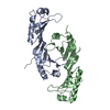
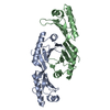
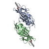
| Entry | Database: PDB / ID: 3eik | ||||||
|---|---|---|---|---|---|---|---|
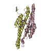
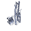
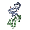
| Title | double stranded DNA binding protein | ||||||
 Components Components | TATA-box-binding protein | ||||||
 Keywords Keywords | TRANSCRIPTION / TATA box binding protein / DNA-binding / Initiation factor / Nucleus | ||||||
| Function / homology |  Function and homology information Function and homology informationtranscription initiation at RNA polymerase II promoter / transcription regulator complex / DNA binding / identical protein binding / nucleus Similarity search - Function | ||||||
| Biological species |  Encephalitozoon cuniculi (fungus) Encephalitozoon cuniculi (fungus) | ||||||
| Method |  X-RAY DIFFRACTION / SYNCHROTRON / MOLECULAR REPLACEMENT / Resolution: 1.9 Å X-RAY DIFFRACTION / SYNCHROTRON / MOLECULAR REPLACEMENT / Resolution: 1.9 Å | ||||||
 Authors Authors | Cui, S. / Wollmann, P. / Moldt, M. / Hopfner, K.-P. | ||||||
 Citation Citation | Journal: To be Published Title: structural studies of ecTBP Authors: Cui, S. / Wollmann, P. / Moldt, M. / Hopfner, K.-P. | ||||||
| History |
|
- Structure visualization
| Structure viewer | Molecule: MolmilJmol/JSmol |
|---|
- Downloads & links
-Download
| PDBx/mmCIF format | 3eik.cif.gz | 166.6 KB | Display | PDBx/mmCIF format |
|---|---|---|---|---|
| PDB format | pdb3eik.ent.gz | 131.6 KB | Display | PDB format |
| PDBx/mmJSON format | 3eik.json.gz | Tree view | PDBx/mmJSON format | |
| Others |  Other downloads Other downloads |
-Validation report
| Arichive directory | https://data.pdbj.org/pub/pdb/validation_reports/ei/3eikftp://data.pdbj.org/pub/pdb/validation_reports/ei/3eik | HTTPS FTP |
|---|
-Related structure data
| Related structure data | 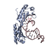 1ytbS S: Starting model for refinement |
|---|---|
| Similar structure data |


-Links
 PDBj
PDBj


- Assembly
| Deposited unit | 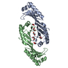
| ||||||||
|---|---|---|---|---|---|---|---|---|---|
| 1 |
| ||||||||
| Unit cell |
|
-Components
| #1: Protein | Mass: 24502.486 Da / Num. of mol.: 2 Source method: isolated from a genetically manipulated source Source: (gene. exp.) Encephalitozoon cuniculi (fungus) / Strain: GB-M1 / Plasmid: pET-28a / Production host:  #2: Chemical | ChemComp-EDO /   Mass: 62.068 Da / Num. of mol.: 6 / Source method: obtained synthetically / Formula: C2H6O2 Mass: 62.068 Da / Num. of mol.: 6 / Source method: obtained synthetically / Formula: C2H6O2#3: Water | ChemComp-HOH / |  Mass: 18.015 Da / Num. of mol.: 148 / Source method: isolated from a natural source / Formula: H2O Mass: 18.015 Da / Num. of mol.: 148 / Source method: isolated from a natural source / Formula: H2O |
|---|
-Experimental details
-Experiment
| Experiment | Method: X-RAY DIFFRACTION / Number of used crystals: 1 |
|---|
- Sample preparation
| Crystal | Density Matthews: 2.76 Å3/Da / Density % sol: 55.47 % |
|---|---|
| Crystal grow | Temperature: 298 K / Method: vapor diffusion, hanging drop / pH: 6.5 Details: 0.1M MES,2M NaCl,4% Acetone, pH 6.5, VAPOR DIFFUSION, HANGING DROP, temperature 298.0K |
-Data collection
| Diffraction | Mean temperature: 100 K |
|---|---|
| Diffraction source | Source: SYNCHROTRON / Site: SLS  / Beamline: X06SA / Wavelength: 1 Å / Beamline: X06SA / Wavelength: 1 Å |
| Detector | Type: PSI PILATUS 6M / Detector: PIXEL / Date: Jun 18, 2008 |
| Radiation | Monochromator: GRAPHITE / Protocol: SINGLE WAVELENGTH / Monochromatic (M) / Laue (L): M / Scattering type: x-ray |
| Radiation wavelength | Wavelength: 1 Å / Relative weight: 1 |
| Reflection | Resolution: 1.9→30 Å / Num. all: 41388 / Num. obs: 37329 / % possible obs: 90.19 % / Observed criterion σ(F): 0 / Observed criterion σ(I): 0 / Redundancy: 3.03 % / Biso Wilson estimate: 25.23 Å2 / Rmerge(I) obs: 0.047 / Rsym value: 0.047 |
| Reflection shell | Resolution: 1.9→2.01 Å / Redundancy: 2.99 % / Rmerge(I) obs: 0.326 / Mean I/σ(I) obs: 3.44 / Num. unique all: 5640 / Rsym value: 0.326 / % possible all: 84.8 |
- Processing
| Software |
| |||||||||||||||||||||||||||||||||||||||||||||||||||||||||||||||||||||||||||||||||||||||||||||||||||||||||
|---|---|---|---|---|---|---|---|---|---|---|---|---|---|---|---|---|---|---|---|---|---|---|---|---|---|---|---|---|---|---|---|---|---|---|---|---|---|---|---|---|---|---|---|---|---|---|---|---|---|---|---|---|---|---|---|---|---|---|---|---|---|---|---|---|---|---|---|---|---|---|---|---|---|---|---|---|---|---|---|---|---|---|---|---|---|---|---|---|---|---|---|---|---|---|---|---|---|---|---|---|---|---|---|---|---|---|
| Refinement | Method to determine structure: MOLECULAR REPLACEMENT Starting model: PDB entry 1YTB Resolution: 1.9→26.1 Å / Occupancy max: 1 / Occupancy min: 0 / FOM work R set: 0.851 / SU ML: 0.31 / σ(F): 1.98 / Stereochemistry target values: ML
| |||||||||||||||||||||||||||||||||||||||||||||||||||||||||||||||||||||||||||||||||||||||||||||||||||||||||
| Solvent computation | Shrinkage radii: 0.9 Å / VDW probe radii: 1.11 Å / Solvent model: FLAT BULK SOLVENT MODEL / Bsol: 54.278 Å2 / ksol: 0.38 e/Å3 | |||||||||||||||||||||||||||||||||||||||||||||||||||||||||||||||||||||||||||||||||||||||||||||||||||||||||
| Displacement parameters | Biso max: 68.87 Å2 / Biso mean: 30.794 Å2 / Biso min: 13.2 Å2
| |||||||||||||||||||||||||||||||||||||||||||||||||||||||||||||||||||||||||||||||||||||||||||||||||||||||||
| Refinement step | Cycle: LAST / Resolution: 1.9→26.1 Å
| |||||||||||||||||||||||||||||||||||||||||||||||||||||||||||||||||||||||||||||||||||||||||||||||||||||||||
| Refine LS restraints |
| |||||||||||||||||||||||||||||||||||||||||||||||||||||||||||||||||||||||||||||||||||||||||||||||||||||||||
| LS refinement shell | Refine-ID: X-RAY DIFFRACTION / Total num. of bins used: 14
|
