- PDB-3eed: Crystal structure of human protein kinase CK2 regulatory subunit ... -
+
Open data
ID or keywords:
Loading...
-
Basic information
Entry
Database: PDB / ID: 3eed
Title
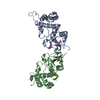
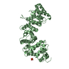
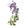
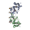
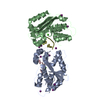
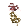
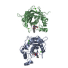
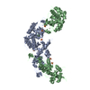
Crystal structure of human protein kinase CK2 regulatory subunit (CK2beta; mutant 1-193)
Components
Casein kinase II subunit beta
Keywords
TRANSFERASE / protein kinase CK2 / casein kinase 2 / casein kinase II / eukaryotic protein kinases / Phosphoprotein / Wnt signaling pathway
Function / homology
Function and homology information
adiponectin-activated signaling pathway / positive regulation of activin receptor signaling pathway / endothelial tube morphogenesis / negative regulation of viral life cycle / protein kinase regulator activity / Phosphorylation and nuclear translocation of BMAL1 (ARNTL) and CLOCK / WNT mediated activation of DVL / Condensation of Prometaphase Chromosomes / protein kinase CK2 complex / symbiont-mediated disruption of host cell PML body ...adiponectin-activated signaling pathway / positive regulation of activin receptor signaling pathway / endothelial tube morphogenesis / negative regulation of viral life cycle / protein kinase regulator activity / Phosphorylation and nuclear translocation of BMAL1 (ARNTL) and CLOCK / WNT mediated activation of DVL / Condensation of Prometaphase Chromosomes / protein kinase CK2 complex / symbiont-mediated disruption of host cell PML body / sperm glycocalyx / Phosphorylation and nuclear translocation of the CRY:PER:kinase complex / perinuclear theca / Regulation of CDH1 posttranslational processing and trafficking to plasma membrane / Receptor Mediated Mitophagy / Synthesis of PC / RUNX1 interacts with co-factors whose precise effect on RUNX1 targets is not known / Maturation of hRSV A proteins / negative regulation of blood vessel endothelial cell migration / positive regulation of SMAD protein signal transduction / negative regulation of proteasomal ubiquitin-dependent protein catabolic process / Signal transduction by L1 / PML body / fibrillar center / Wnt signaling pathway / Regulation of PTEN stability and activity / KEAP1-NFE2L2 pathway / Cooperation of PDCL (PhLP1) and TRiC/CCT in G-protein beta folding / protein-containing complex assembly / sperm midpiece / secretory granule lumen / Regulation of TP53 Activity through Phosphorylation / ficolin-1-rich granule lumen / RNA polymerase II-specific DNA-binding transcription factor binding / protein-macromolecule adaptor activity / signaling receptor binding / negative regulation of cell population proliferation / protein domain specific binding / protein serine/threonine kinase activity / chromatin binding / Neutrophil degranulation / chromatin / signal transduction / extracellular exosome / extracellular region / nucleoplasm / metal ion binding / identical protein binding / nucleus / cytoplasm / cytosol Similarity search - Function
protein kinase ck2 holoenzyme, chain C, domain 1 / protein kinase ck2 holoenzyme, chain C, domain 1 / N-terminal domain of TfIIb - #20 / Casein kinase II, regulatory subunit / Casein kinase II, regulatory subunit, N-terminal / Casein kinase II subunit beta-like / Casein kinase II regulatory subunit / Casein kinase II regulatory subunit signature. / Casein kinase II regulatory subunit / N-terminal domain of TfIIb ...protein kinase ck2 holoenzyme, chain C, domain 1 / protein kinase ck2 holoenzyme, chain C, domain 1 / N-terminal domain of TfIIb - #20 / Casein kinase II, regulatory subunit / Casein kinase II, regulatory subunit, N-terminal / Casein kinase II subunit beta-like / Casein kinase II regulatory subunit / Casein kinase II regulatory subunit signature. / Casein kinase II regulatory subunit / N-terminal domain of TfIIb / Single Sheet / Orthogonal Bundle / Mainly Beta / Mainly Alpha Similarity search - Domain/homology
Resolution: 2.8→29.12 Å / Cor.coef. Fo:Fc: 0.957 / Cor.coef. Fo:Fc free: 0.934 / SU B: 22.332 / SU ML: 0.221 / Cross valid method: THROUGHOUT / σ(F): 0 / ESU R Free: 0.318 / Stereochemistry target values: MAXIMUM LIKELIHOOD / Details: HYDROGENS HAVE BEEN ADDED IN THE RIDING POSITIONS
Rfactor
Num. reflection
% reflection
Selection details
Rfree
0.21616
1321
9.9 %
RANDOM
Rwork
0.17163
-
-
-
all
0.17619
12061
-
-
obs
0.17619
12061
100 %
-
Solvent computation
Ion probe radii: 0.8 Å / Shrinkage radii: 0.8 Å / VDW probe radii: 1.2 Å / Solvent model: BABINET MODEL WITH MASK
Displacement parameters
Biso mean: 52.049 Å2
Baniso -1
Baniso -2
Baniso -3
1-
0.47 Å2
0 Å2
0 Å2
2-
-
0.47 Å2
0 Å2
3-
-
-
-0.94 Å2
Refinement step
Cycle: LAST / Resolution: 2.8→29.12 Å
Protein
Nucleic acid
Ligand
Solvent
Total
Num. atoms
3000
0
17
82
3099
Refine LS restraints
Refine-ID
Type
Dev ideal
Dev ideal target
Number
X-RAY DIFFRACTION
r_bond_refined_d
0.011
0.022
3112
X-RAY DIFFRACTION
r_bond_other_d
0.001
0.02
2140
X-RAY DIFFRACTION
r_angle_refined_deg
1.123
1.961
4216
X-RAY DIFFRACTION
r_angle_other_deg
0.736
3.003
5170
X-RAY DIFFRACTION
r_dihedral_angle_1_deg
5.685
5
362
X-RAY DIFFRACTION
r_dihedral_angle_2_deg
34.365
23.902
164
X-RAY DIFFRACTION
r_dihedral_angle_3_deg
14.482
15
502
X-RAY DIFFRACTION
r_dihedral_angle_4_deg
20.896
15
18
X-RAY DIFFRACTION
r_chiral_restr
0.059
0.2
418
X-RAY DIFFRACTION
r_gen_planes_refined
0.006
0.021
3456
X-RAY DIFFRACTION
r_gen_planes_other
0.002
0.02
654
X-RAY DIFFRACTION
r_nbd_refined
X-RAY DIFFRACTION
r_nbd_other
X-RAY DIFFRACTION
r_nbtor_refined
X-RAY DIFFRACTION
r_nbtor_other
X-RAY DIFFRACTION
r_xyhbond_nbd_refined
X-RAY DIFFRACTION
r_xyhbond_nbd_other
X-RAY DIFFRACTION
r_metal_ion_refined
X-RAY DIFFRACTION
r_metal_ion_other
X-RAY DIFFRACTION
r_symmetry_vdw_refined
X-RAY DIFFRACTION
r_symmetry_vdw_other
X-RAY DIFFRACTION
r_symmetry_hbond_refined
X-RAY DIFFRACTION
r_symmetry_hbond_other
X-RAY DIFFRACTION
r_symmetry_metal_ion_refined
X-RAY DIFFRACTION
r_symmetry_metal_ion_other
X-RAY DIFFRACTION
r_mcbond_it
2.25
6
1830
X-RAY DIFFRACTION
r_mcbond_other
0.538
6
728
X-RAY DIFFRACTION
r_mcangle_it
3.652
9
2956
X-RAY DIFFRACTION
r_scbond_it
3.146
6
1282
X-RAY DIFFRACTION
r_scangle_it
4.387
9
1260
X-RAY DIFFRACTION
r_rigid_bond_restr
X-RAY DIFFRACTION
r_sphericity_free
X-RAY DIFFRACTION
r_sphericity_bonded
Refine LS restraints NCS
Dom-ID: 1 / Refine-ID: X-RAY DIFFRACTION
Ens-ID
Auth asym-ID
Number
Type
Rms dev position (Å)
Weight position
1
A
296
TIGHTPOSITIONAL
0.03
0.05
1
A
409
MEDIUMPOSITIONAL
0.03
0.5
1
B
296
TIGHTTHERMAL
0.08
0.5
1
B
409
MEDIUMTHERMAL
0.08
2
2
A
729
TIGHTPOSITIONAL
0.03
0.05
2
A
1026
MEDIUMPOSITIONAL
0.04
0.5
2
B
729
TIGHTTHERMAL
0.09
0.5
2
B
1026
MEDIUMTHERMAL
0.09
2
LS refinement shell
Resolution: 2.8→2.872 Å / Total num. of bins used: 20
Rfactor
Num. reflection
% reflection
Rfree
0.372
74
-
Rwork
0.292
880
-
obs
-
-
100 %
Refinement TLS params.
Method: refined / Refine-ID: X-RAY DIFFRACTION
ID
L11 (°2)
L12 (°2)
L13 (°2)
L22 (°2)
L23 (°2)
L33 (°2)
S11 (Å °)
S12 (Å °)
S13 (Å °)
S21 (Å °)
S22 (Å °)
S23 (Å °)
S31 (Å °)
S32 (Å °)
S33 (Å °)
T11 (Å2)
T12 (Å2)
T13 (Å2)
T22 (Å2)
T23 (Å2)
T33 (Å2)
Origin x (Å)
Origin y (Å)
Origin z (Å)
1
2.4177
0.0966
-0.2228
4.2544
1.6331
5.1547
0.0372
0.1722
-0.2837
0.0767
0.0202
-0.4449
0.4873
0.5517
-0.0574
-0.1598
0.101
-0.0582
-0.125
0.0167
-0.0777
-19.31
-34.262
-25.498
2
1.1223
0.877
1.3683
5.0304
1.93
3.4109
0.1794
-0.0162
-0.0982
0.5219
-0.2045
0.1873
0.1992
-0.1768
0.0251
-0.1474
-0.0321
0
-0.196
0.0252
-0.2155
-28.535
-18.419
-14.462
3
1.4103
-4.4601
0.1116
35.3621
7.0648
8.0381
-0.4561
0.4334
0.4656
0.9354
0.6746
0.6009
-0.1427
-1.1647
-0.2186
-0.1373
-0.0134
0.1084
0.0498
0.0033
-0.1165
-33.719
7.302
-1.846
4
3.0115
0.6706
1.842
3.7388
-0.2804
5.9776
-0.0596
-0.3595
0.0572
0.7412
-0.0148
-0.6397
-0.3347
0.8772
0.0743
-0.03
-0.0759
-0.0949
0.0943
-0.0002
-0.0325
-8.727
12.526
1.53
5
2.1987
0.7103
0.3821
4.9288
0.9461
3.1904
0.0076
-0.049
0.0195
0.0209
-0.0025
0.0429
0.1275
-0.0448
-0.005
-0.2284
0.0103
0.0411
-0.2325
0.0425
-0.1877
-23.583
2.53
-10.242
6
12.0995
17.8715
1.139
26.619
2.7172
4.9271
-0.831
0.369
0.4486
-1.4287
0.9275
0.4757
-0.0528
-0.5749
-0.0964
-0.1746
0.0089
0.0158
-0.0843
-0.0245
-0.0464
-39.393
-18.085
-23.608
Refinement TLS group
ID
Refine-ID
Refine TLS-ID
Auth asym-ID
Label asym-ID
Auth seq-ID
Label seq-ID
1
X-RAY DIFFRACTION
1
A
A
6 - 59
6 - 59
2
X-RAY DIFFRACTION
1
A
A
65 - 105
65 - 105
3
X-RAY DIFFRACTION
2
A
A
106 - 180
106 - 180
4
X-RAY DIFFRACTION
3
A
A
181 - 193
181 - 193
5
X-RAY DIFFRACTION
4
B
B
6 - 59
6 - 59
6
X-RAY DIFFRACTION
4
B
B
65 - 105
65 - 105
7
X-RAY DIFFRACTION
5
B
B
106 - 180
106 - 180
8
X-RAY DIFFRACTION
6
B
B
181 - 193
181 - 193
+
About Yorodumi
-
News
-
Feb 9, 2022. New format data for meta-information of EMDB entries
New format data for meta-information of EMDB entries
Version 3 of the EMDB header file is now the official format.
The previous official version 1.9 will be removed from the archive.
In the structure databanks used in Yorodumi, some data are registered as the other names, "COVID-19 virus" and "2019-nCoV". Here are the details of the virus and the list of structure data.
Jan 31, 2019. EMDB accession codes are about to change! (news from PDBe EMDB page)
EMDB accession codes are about to change! (news from PDBe EMDB page)
The allocation of 4 digits for EMDB accession codes will soon come to an end. Whilst these codes will remain in use, new EMDB accession codes will include an additional digit and will expand incrementally as the available range of codes is exhausted. The current 4-digit format prefixed with “EMD-” (i.e. EMD-XXXX) will advance to a 5-digit format (i.e. EMD-XXXXX), and so on. It is currently estimated that the 4-digit codes will be depleted around Spring 2019, at which point the 5-digit format will come into force.
The EM Navigator/Yorodumi systems omit the EMD- prefix.
Related info.:Q: What is EMD? / ID/Accession-code notation in Yorodumi/EM Navigator
Yorodumi is a browser for structure data from EMDB, PDB, SASBDB, etc.
This page is also the successor to EM Navigator detail page, and also detail information page/front-end page for Omokage search.
The word "yorodu" (or yorozu) is an old Japanese word meaning "ten thousand". "mi" (miru) is to see.
Related info.:EMDB / PDB / SASBDB / Comparison of 3 databanks / Yorodumi Search / Aug 31, 2016. New EM Navigator & Yorodumi / Yorodumi Papers / Jmol/JSmol / Function and homology information / Changes in new EM Navigator and Yorodumi
 Movie
Movie Controller

 Yorodumi
 Open data
 Basic information
 Components
 Keywords
 Function and homology information
 Homo sapiens (human)
 X-RAY DIFFRACTION /
  Authors
 Citation
 Structure visualization
 Downloads & links
 Other downloads


 PDBj
PDBj


 Assembly


 Mass: 65.409 Da / Num. of mol.: 2 / Source method: obtained synthetically / Formula: Zn
 Mass: 96.063 Da / Num. of mol.: 3 / Source method: obtained synthetically / Formula: SO4
 Mass: 18.015 Da / Num. of mol.: 82 / Source method: isolated from a natural source / Formula: H2O
 Sample preparation
 / Beamline: X12 / Wavelength: 0.9 Å
 Processing