 Movie
Movie Controller

[English] 日本語
 Yorodumi
- PDB-3eb2: Crystal structure of Dihydrodipicolinate Synthase from Rhodopseud... -
+ Open data

- Basic information
| Entry | Database: PDB / ID: 3eb2 | ||||||
|---|---|---|---|---|---|---|---|
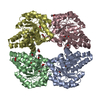
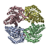
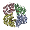
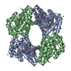
| Title | Crystal structure of Dihydrodipicolinate Synthase from Rhodopseudomonas palustris at 2.0A resolution | ||||||
 Components Components | Putative dihydrodipicolinate synthetase | ||||||
 Keywords Keywords | LYASE / LYSINE BIOSYNTHESIS / PYRUVATE / TIM BARREL / NYSGXRC / 11102O / PSI2. / Structural Genomics / Protein Structure Initiative / New York SGX Research Center for Structural Genomics | ||||||
| Function / homology |  Function and homology information Function and homology informationdihydrodipicolinate synthase / 4-hydroxy-tetrahydrodipicolinate synthase activity Similarity search - Function | ||||||
| Biological species |  Rhodopseudomonas palustris (phototrophic) Rhodopseudomonas palustris (phototrophic) | ||||||
| Method |  X-RAY DIFFRACTION / SYNCHROTRON / SAD / Resolution: 2.04 Å X-RAY DIFFRACTION / SYNCHROTRON / SAD / Resolution: 2.04 Å | ||||||
 Authors Authors | Satyanarayana, L. / Eswaramoorthy, S. / Burley, S.K. / Swaminathan, S. / New York SGX Research Center for Structural Genomics (NYSGXRC) | ||||||
 Citation Citation | Journal: To be Published Title: Crystal structure of Dihydrodipicolinate Synthase from Rhodopseudomonas palustris at 2.0A resolution Authors: Satyanarayana, L. / Eswaramoorthy, S. / Burley, S.K. / Swaminathan, S. | ||||||
| History |
|
- Structure visualization
| Structure viewer | Molecule: MolmilJmol/JSmol |
|---|
- Downloads & links
-Download
| PDBx/mmCIF format | 3eb2.cif.gz | 233.4 KB | Display | PDBx/mmCIF format |
|---|---|---|---|---|
| PDB format | pdb3eb2.ent.gz | 188 KB | Display | PDB format |
| PDBx/mmJSON format | 3eb2.json.gz | Tree view | PDBx/mmJSON format | |
| Others |  Other downloads Other downloads |
-Validation report
| Arichive directory | https://data.pdbj.org/pub/pdb/validation_reports/eb/3eb2ftp://data.pdbj.org/pub/pdb/validation_reports/eb/3eb2 | HTTPS FTP |
|---|
-Related structure data
| Related structure data | |
|---|---|
| Similar structure data | |
| Other databases |


-Links
 PDBj
PDBj- Assembly
| Deposited unit | 
| ||||||||
|---|---|---|---|---|---|---|---|---|---|
| 1 |
| ||||||||
| Unit cell |
|
-Components
| #1: Protein | Mass: 32508.338 Da / Num. of mol.: 4 Source method: isolated from a genetically manipulated source Details: Top10-Invitrogen Source: (gene. exp.) Rhodopseudomonas palustris (phototrophic)Strain: palustris / Gene: dapA1, RPA1571 / Plasmid: BC-pSGX3(BC) / Production host: #2: Chemical |   Mass: 150.173 Da / Num. of mol.: 2 / Source method: obtained synthetically / Formula: C6H14O4 Mass: 150.173 Da / Num. of mol.: 2 / Source method: obtained synthetically / Formula: C6H14O4#3: Water | ChemComp-HOH / |  Mass: 18.015 Da / Num. of mol.: 605 / Source method: isolated from a natural source / Formula: H2O Mass: 18.015 Da / Num. of mol.: 605 / Source method: isolated from a natural source / Formula: H2O |
|---|
-Experimental details
-Experiment
| Experiment | Method: X-RAY DIFFRACTION / Number of used crystals: 1 |
|---|
- Sample preparation
| Crystal | Density Matthews: 2.5 Å3/Da / Density % sol: 50.85 % |
|---|---|
| Crystal grow | Method: vapor diffusion, sitting drop / pH: 5.5 Details: 25% PEG 1,500, 0.1M Bis-Tris pH 5.5, 0.025M Trimethylamine hydrochloride., VAPOR DIFFUSION, SITTING DROP |
-Data collection
| Diffraction | Mean temperature: 100 K |
|---|---|
| Diffraction source | Source: SYNCHROTRON / Site: NSLS  / Beamline: X12C / Wavelength: 0.98 Å / Beamline: X12C / Wavelength: 0.98 Å |
| Detector | Type: ADSC QUANTUM 210 / Detector: CCD / Date: Aug 10, 2008 / Details: mirrors |
| Radiation | Monochromator: Si 111 CHANNEL / Protocol: SINGLE WAVELENGTH / Monochromatic (M) / Laue (L): M / Scattering type: x-ray |
| Radiation wavelength | Wavelength: 0.98 Å / Relative weight: 1 |
| Reflection | Resolution: 2.04→50 Å / Num. all: 82938 / Num. obs: 82938 / % possible obs: 97.9 % / Observed criterion σ(F): 0 / Observed criterion σ(I): 0 / Redundancy: 12.9 % / Biso Wilson estimate: 13.5 Å2 / Rmerge(I) obs: 0.086 / Net I/σ(I): 13.8 |
| Reflection shell | Resolution: 2.04→2.11 Å / Redundancy: 10.2 % / Rmerge(I) obs: 0.433 / Mean I/σ(I) obs: 11.1 / Num. unique all: 7055 / % possible all: 84.3 |
- Processing
| Software |
| |||||||||||||||||||||||||
|---|---|---|---|---|---|---|---|---|---|---|---|---|---|---|---|---|---|---|---|---|---|---|---|---|---|---|
| Refinement | Method to determine structure: SAD / Resolution: 2.04→50 Å / Isotropic thermal model: Isotropic / Cross valid method: THROUGHOUT / σ(F): 0 / σ(I): 0 / Stereochemistry target values: Engh & Huber
| |||||||||||||||||||||||||
| Displacement parameters | Biso mean: 25.3 Å2
| |||||||||||||||||||||||||
| Refine analyze |
| |||||||||||||||||||||||||
| Refinement step | Cycle: LAST / Resolution: 2.04→50 Å
| |||||||||||||||||||||||||
| Refine LS restraints |
| |||||||||||||||||||||||||
| LS refinement shell | Resolution: 2.04→2.17 Å / Rfactor Rfree error: 0.013
|
