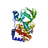
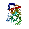
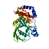
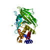
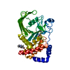
Entry Database : PDB / ID : 3eaxTitle Crystal structure PTP1B complex with small molecule compound LZP-6 Tyrosine-protein phosphatase non-receptor type 1 Keywords / / / / / / / / / / Function / homology Function Domain/homology Component
/ / / / / / / / / / / / / / / / / / / / / / / / / / / / / / / / / / / / / / / / / / / / / / / / / / / / / / / / / / / / / / / / / / / / / / / / / / / / / / / / / / / / / / / / / / / / / / / / / / / / / / / / / / / / / / Biological species Homo sapiens (human)Method / / Resolution : 1.9 Å Authors Zhang, Z.-Y. / Liu, S. / Zhang, L.-F. / Yu, X. / Xue, T. / Gunawan, A.M. / Long, Y.-Q. Journal : J.Am.Chem.Soc. / Year : 2008Title : Targeting inactive enzyme conformation: aryl diketoacid derivatives as a new class of PTP1B inhibitors.Authors : Liu, S. / Zeng, L.F. / Wu, L. / Yu, X. / Xue, T. / Gunawan, A.M. / Long, Y.Q. / Zhang, Z.Y. History Deposition Aug 26, 2008 Deposition site / Processing site Revision 1.0 Jul 7, 2009 Provider / Type Revision 1.1 Jul 13, 2011 Group Revision 1.2 Feb 21, 2024 Group / Database references / Derived calculationsCategory chem_comp_atom / chem_comp_bond ... chem_comp_atom / chem_comp_bond / database_2 / struct_site Item _database_2.pdbx_DOI / _database_2.pdbx_database_accession ... _database_2.pdbx_DOI / _database_2.pdbx_database_accession / _struct_site.pdbx_auth_asym_id / _struct_site.pdbx_auth_comp_id / _struct_site.pdbx_auth_seq_id
Show all Show less
 Movie
Movie Controller

 Yorodumi
 Open data
 Basic information
 Components
 Keywords
 Function and homology information
 Homo sapiens (human)
 X-RAY DIFFRACTION /
  Authors
 Citation
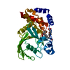
 Structure visualization
 Downloads & links
 Other downloads
 PDBj
PDBj


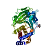
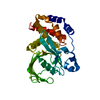
 Assembly
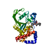


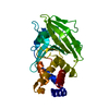
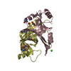
 Mass: 646.685 Da / Num. of mol.: 1 / Source method: obtained synthetically / Formula: C38H34N2O8
 Mass: 18.015 Da / Num. of mol.: 90 / Source method: isolated from a natural source / Formula: H2O
 Sample preparation
 Processing