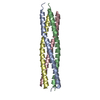
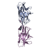
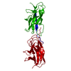
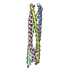
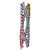
- PDB-3e7k: Crystal Structure of an antiparallel coiled-coil tetramerization ... -
+
Open data
ID or keywords:
Loading...
-
Basic information
Entry
Database: PDB / ID: 3e7k
Title
Crystal Structure of an antiparallel coiled-coil tetramerization domain from TRPM7 channels
Components
TRPM7 channel
Keywords
MEMBRANE PROTEIN / Coiled-coil / antiparallel / Ion Channel / Assembly domain / TRPM channel / TRPM7
Function / homology
Function and homology information
calcium-dependent cell-matrix adhesion / intracellular magnesium ion homeostasis / varicosity / magnesium ion transport / magnesium ion transmembrane transport / magnesium ion homeostasis / zinc ion transport / zinc ion transmembrane transporter activity / magnesium ion transmembrane transporter activity / TRP channels ...calcium-dependent cell-matrix adhesion / intracellular magnesium ion homeostasis / varicosity / magnesium ion transport / magnesium ion transmembrane transport / magnesium ion homeostasis / zinc ion transport / zinc ion transmembrane transporter activity / magnesium ion transmembrane transporter activity / TRP channels / actomyosin structure organization / myosin binding / necroptotic process / monoatomic cation transmembrane transport / monoatomic cation channel activity / ruffle / calcium ion transmembrane transport / calcium channel activity / memory / kinase activity / calcium ion transport / synaptic vesicle membrane / actin binding / cytoplasmic vesicle / protein homotetramerization / protein kinase activity / non-specific serine/threonine protein kinase / positive regulation of apoptotic process / protein serine kinase activity / protein serine/threonine kinase activity / neuronal cell body / ATP binding / metal ion binding / nucleus / plasma membrane Similarity search - Function
TRPM, tetramerisation domain / Transient receptor potential cation channel subfamily M member 7 / MHCK/EF2 kinase / Alpha-kinase family / Alpha-type protein kinase domain profile. / Alpha-kinase family / TRPM, tetramerisation domain / TRPM, tetramerisation domain superfamily / Tetramerisation domain of TRPM / TRPM, SLOG domain ...TRPM, tetramerisation domain / Transient receptor potential cation channel subfamily M member 7 / MHCK/EF2 kinase / Alpha-kinase family / Alpha-type protein kinase domain profile. / Alpha-kinase family / TRPM, tetramerisation domain / TRPM, tetramerisation domain superfamily / Tetramerisation domain of TRPM / TRPM, SLOG domain / : / : / SLOG in TRPM / TRPM2-like domain / Single alpha-helices involved in coiled-coils or other helix-helix interfaces / Ion transport domain / Ion transport protein / Protein kinase-like domain superfamily / Up-down Bundle / Mainly Alpha Similarity search - Domain/homology
In the structure databanks used in Yorodumi, some data are registered as the other names, "COVID-19 virus" and "2019-nCoV". Here are the details of the virus and the list of structure data.
Jan 31, 2019. EMDB accession codes are about to change! (news from PDBe EMDB page)
EMDB accession codes are about to change! (news from PDBe EMDB page)
The allocation of 4 digits for EMDB accession codes will soon come to an end. Whilst these codes will remain in use, new EMDB accession codes will include an additional digit and will expand incrementally as the available range of codes is exhausted. The current 4-digit format prefixed with “EMD-” (i.e. EMD-XXXX) will advance to a 5-digit format (i.e. EMD-XXXXX), and so on. It is currently estimated that the 4-digit codes will be depleted around Spring 2019, at which point the 5-digit format will come into force.
The EM Navigator/Yorodumi systems omit the EMD- prefix.
Related info.:Q: What is EMD? / ID/Accession-code notation in Yorodumi/EM Navigator
Yorodumi is a browser for structure data from EMDB, PDB, SASBDB, etc.
This page is also the successor to EM Navigator detail page, and also detail information page/front-end page for Omokage search.
The word "yorodu" (or yorozu) is an old Japanese word meaning "ten thousand". "mi" (miru) is to see.
Related info.:EMDB / PDB / SASBDB / Comparison of 3 databanks / Yorodumi Search / Aug 31, 2016. New EM Navigator & Yorodumi / Yorodumi Papers / Jmol/JSmol / Function and homology information / Changes in new EM Navigator and Yorodumi
 Movie
Movie Controller

 Yorodumi
 Open data
 Basic information
 Components
 Keywords
 Function and homology information
 X-RAY DIFFRACTION /
  Authors
 Citation
 Structure visualization
 Downloads & links
 Other downloads
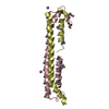


 PDBj
PDBj
 Assembly

 Mass: 18.015 Da / Num. of mol.: 296 / Source method: isolated from a natural source / Formula: H2O
 Sample preparation
 / Beamline: BL9-2 / Wavelength: 0.9796 Å
 Processing