 Movie
Movie Controller

[English] 日本語
 Yorodumi
- PDB-1iam: STRUCTURE OF THE TWO AMINO-TERMINAL DOMAINS OF HUMAN INTERCELLULA... -
+ Open data

- Basic information
| Entry | Database: PDB / ID: 1iam | ||||||
|---|---|---|---|---|---|---|---|
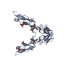
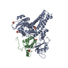
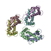
| Title | STRUCTURE OF THE TWO AMINO-TERMINAL DOMAINS OF HUMAN INTERCELLULAR ADHESION MOLECULE-1, ICAM-1 | ||||||
 Components Components | INTERCELLULAR ADHESION MOLECULE-1 | ||||||
 Keywords Keywords | Viral protein receptor / RHINOVIRUS RECEPTOR / CELL ADHESION / INTEGRIN LIGAND / GLYCOPROTEIN / LFA-1 LIGAND / IMMUNOGLOBULIN FOLD / TRANSMEMBRANE | ||||||
| Function / homology |  Function and homology information Function and homology informationregulation of leukocyte mediated cytotoxicity / T cell extravasation / positive regulation of cellular extravasation / leukocyte adhesion to vascular endothelial cell / regulation of ruffle assembly / membrane to membrane docking / T cell antigen processing and presentation / cell-cell adhesion mediated by integrin / T cell activation via T cell receptor contact with antigen bound to MHC molecule on antigen presenting cell / adhesion of symbiont to host ...regulation of leukocyte mediated cytotoxicity / T cell extravasation / positive regulation of cellular extravasation / leukocyte adhesion to vascular endothelial cell / regulation of ruffle assembly / membrane to membrane docking / T cell antigen processing and presentation / cell-cell adhesion mediated by integrin / T cell activation via T cell receptor contact with antigen bound to MHC molecule on antigen presenting cell / adhesion of symbiont to host / establishment of endothelial barrier / heterophilic cell-cell adhesion / leukocyte migration / leukocyte cell-cell adhesion / Interleukin-10 signaling / immunological synapse / Integrin cell surface interactions / negative regulation of endothelial cell apoptotic process / negative regulation of extrinsic apoptotic signaling pathway via death domain receptors / cellular response to leukemia inhibitory factor / cellular response to glucose stimulus / integrin binding / cellular response to amyloid-beta / Interferon gamma signaling / Immunoregulatory interactions between a Lymphoid and a non-Lymphoid cell / transmembrane signaling receptor activity / virus receptor activity / extracellular matrix / signaling receptor activity / Interleukin-4 and Interleukin-13 signaling / receptor-mediated virion attachment to host cell / positive regulation of ERK1 and ERK2 cascade / cell adhesion / membrane raft / receptor ligand activity / external side of plasma membrane / focal adhesion / cell surface / : / extracellular exosome / membrane / plasma membrane Similarity search - Function | ||||||
| Biological species |  Homo sapiens (human) Homo sapiens (human) | ||||||
| Method |  X-RAY DIFFRACTION / SYNCHROTRON / MOLECULAR REPLACEMENT, MIR, PHASE RECOMBINATION / Resolution: 2.1 Å X-RAY DIFFRACTION / SYNCHROTRON / MOLECULAR REPLACEMENT, MIR, PHASE RECOMBINATION / Resolution: 2.1 Å | ||||||
 Authors Authors | Bella, J. / Kolatkar, P.R. / Marlor, C. / Greve, J.M. / Rossmann, M.G. | ||||||
 Citation Citation | Journal: Proc.Natl.Acad.Sci.USA / Year: 1998 Title: The structure of the two amino-terminal domains of human ICAM-1 suggests how it functions as a rhinovirus receptor and as an LFA-1 integrin ligand. Authors: Bella, J. / Kolatkar, P.R. / Marlor, C.W. / Greve, J.M. / Rossmann, M.G. #1: Journal: Proc.Natl.Acad.Sci.USA / Year: 1993Title: Structure of a Human Rhinovirus Complexed with its Receptor Molecule Authors: Olson, N.H. / Kolatkar, P.R. / Oliveira, M.A. / Cheng, R.H. / Greve, J.M. / Mcclelland, A. / Baker, T.S. / Rossmann, M.G. #2: Journal: Cell(Cambridge,Mass.) / Year: 1989Title: The Major Human Rhinovirus Receptor is Icam-1 Authors: Greve, J.M. / Davis, G. / Meyer, A.M. / Forte, C.P. / Yost, S.C. / Marlor, C.W. / Kamarck, M.E. / Mcclelland, A. | ||||||
| History |
|
- Structure visualization
| Structure viewer | Molecule: MolmilJmol/JSmol |
|---|
- Downloads & links
-Download
| PDBx/mmCIF format | 1iam.cif.gz | 53.9 KB | Display | PDBx/mmCIF format |
|---|---|---|---|---|
| PDB format | pdb1iam.ent.gz | 37.8 KB | Display | PDB format |
| PDBx/mmJSON format | 1iam.json.gz | Tree view | PDBx/mmJSON format | |
| Others |  Other downloads Other downloads |
-Validation report
| Arichive directory | https://data.pdbj.org/pub/pdb/validation_reports/ia/1iamftp://data.pdbj.org/pub/pdb/validation_reports/ia/1iam | HTTPS FTP |
|---|
-Related structure data
| Related structure data |  1zxqS S: Starting model for refinement |
|---|---|
| Similar structure data |


-Links
 PDBj
PDBj


- Assembly
| Deposited unit | 
| |||||||||
|---|---|---|---|---|---|---|---|---|---|---|
| 1 | 
| |||||||||
| Unit cell |
| |||||||||
| Components on special symmetry positions |
|
-Components
| #1: Protein | Mass: 20480.338 Da / Num. of mol.: 1 / Fragment: TWO N-TERMINAL, IMMUNOGLOBULIN DOMAINS / Mutation: N103Q, N118Q, N156Q Source method: isolated from a genetically manipulated source Source: (gene. exp.) Homo sapiens (human) / Cell: LEUKOCYTES, ENDOTHELIAL CELLS / Cell line: SF9 / Plasmid: PVL1393 (PHARMINGEN) / Cell line (production host): SF9 / Production host:   Spodoptera frugiperda (fall armyworm) / References: UniProt: P05362 Spodoptera frugiperda (fall armyworm) / References: UniProt: P05362 |
|---|---|
| #2: Sugar | ChemComp-NAG /   Type: D-saccharide, beta linking / Mass: 221.208 Da / Num. of mol.: 1 Type: D-saccharide, beta linking / Mass: 221.208 Da / Num. of mol.: 1Source method: isolated from a genetically manipulated source Formula: C8H15NO6 |
| #3: Water | ChemComp-HOH /  Mass: 18.015 Da / Num. of mol.: 147 / Source method: isolated from a natural source / Formula: H2O Mass: 18.015 Da / Num. of mol.: 147 / Source method: isolated from a natural source / Formula: H2O |
| Has protein modification | Y |
-Experimental details
-Experiment
| Experiment | Method: X-RAY DIFFRACTION / Number of used crystals: 1 |
|---|
- Sample preparation
| Crystal | Density Matthews: 2.65 Å3/Da / Density % sol: 53.56 % Description: THERE IS A SIGNIFICANT NUMBER OF OVERLOADS IN THE LOWER RESOLUTION SHELLS (19% BETWEEN 20 AN 5 ANGSTROM). DATA WAS SCALED WITH THE "INCLUDE OVERLOADS" OPTION IN SCALEPACK. LOW RESOLUTION ...Description: THERE IS A SIGNIFICANT NUMBER OF OVERLOADS IN THE LOWER RESOLUTION SHELLS (19% BETWEEN 20 AN 5 ANGSTROM). DATA WAS SCALED WITH THE "INCLUDE OVERLOADS" OPTION IN SCALEPACK. LOW RESOLUTION DATA BETWEEN 20 AND 5 ANGSTROM HAS NOT BEEN USED IN THE REFINEMENT. | ||||||||||||||||||||||||||||||||||||
|---|---|---|---|---|---|---|---|---|---|---|---|---|---|---|---|---|---|---|---|---|---|---|---|---|---|---|---|---|---|---|---|---|---|---|---|---|---|
| Crystal grow | pH: 7.5 Details: PROTEIN IN 10 MM TRIS, PH 7.5, 25 MM NACL, WAS CRYSTALLIZED FROM 20% PEG 4000 IN 10 MM TRIS AS PRECIPITANT | ||||||||||||||||||||||||||||||||||||
| Crystal grow | *PLUS Method: vapor diffusion, hanging drop | ||||||||||||||||||||||||||||||||||||
| Components of the solutions | *PLUS
|
-Data collection
| Diffraction | Mean temperature: 120 K |
|---|---|
| Diffraction source | Source: SYNCHROTRON / Site: CHESS  / Beamline: A1 / Wavelength: 0.92 / Beamline: A1 / Wavelength: 0.92 |
| Detector | Detector: CCD / Date: Feb 26, 1995 |
| Radiation | Monochromatic (M) / Laue (L): M / Scattering type: x-ray |
| Radiation wavelength | Wavelength: 0.92 Å / Relative weight: 1 |
| Reflection | Resolution: 2.1→20 Å / Num. obs: 10629 / % possible obs: 81.1 % / Observed criterion σ(I): 2 / Redundancy: 3 % / Biso Wilson estimate: 25.1 Å2 / Rmerge(I) obs: 0.036 / Net I/σ(I): 24.15 |
| Reflection shell | Resolution: 2.1→2.17 Å / Redundancy: 0.3 % / Rmerge(I) obs: 0.059 / Mean I/σ(I) obs: 10.21 / % possible all: 33.4 |
- Processing
| Software |
| ||||||||||||||||||||||||||||||||||||||||||||||||||||||||||||||||||||||||||||||||
|---|---|---|---|---|---|---|---|---|---|---|---|---|---|---|---|---|---|---|---|---|---|---|---|---|---|---|---|---|---|---|---|---|---|---|---|---|---|---|---|---|---|---|---|---|---|---|---|---|---|---|---|---|---|---|---|---|---|---|---|---|---|---|---|---|---|---|---|---|---|---|---|---|---|---|---|---|---|---|---|---|---|
| Refinement | Method to determine structure: MOLECULAR REPLACEMENT, MIR, PHASE RECOMBINATION Starting model: ICAM-2, PDB ENTRY 1ZXQ Resolution: 2.1→5 Å / Rfactor Rfree error: 0.01 / Data cutoff high absF: 100000 / Data cutoff low absF: 0.001 / Isotropic thermal model: RESTRAINED / Cross valid method: THROUGHOUT / σ(F): 0
| ||||||||||||||||||||||||||||||||||||||||||||||||||||||||||||||||||||||||||||||||
| Displacement parameters | Biso mean: 24.6 Å2
| ||||||||||||||||||||||||||||||||||||||||||||||||||||||||||||||||||||||||||||||||
| Refinement step | Cycle: LAST / Resolution: 2.1→5 Å
| ||||||||||||||||||||||||||||||||||||||||||||||||||||||||||||||||||||||||||||||||
| Refine LS restraints |
| ||||||||||||||||||||||||||||||||||||||||||||||||||||||||||||||||||||||||||||||||
| LS refinement shell | Resolution: 2.1→2.19 Å / Rfactor Rfree error: 0.037 / Total num. of bins used: 8
| ||||||||||||||||||||||||||||||||||||||||||||||||||||||||||||||||||||||||||||||||
| Xplor file |
| ||||||||||||||||||||||||||||||||||||||||||||||||||||||||||||||||||||||||||||||||
| Software | *PLUS Name: X-PLOR / Version: 3.851 / Classification: refinement | ||||||||||||||||||||||||||||||||||||||||||||||||||||||||||||||||||||||||||||||||
| Refine LS restraints | *PLUS
|
