 Movie
Movie Controller

[English] 日本語
 Yorodumi
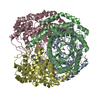
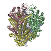
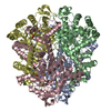
- PDB-3e5b: 2.4 A crystal structure of isocitrate lyase from brucella melitensis -
+ Open data

- Basic information
| Entry | Database: PDB / ID: 3e5b | ||||||
|---|---|---|---|---|---|---|---|
| Title | 2.4 A crystal structure of isocitrate lyase from brucella melitensis | ||||||
 Components Components | isocitrate lyase | ||||||
 Keywords Keywords | LYASE / BRUCELLA / MELITENSIS / ISOCITRATE / SEATTLE STRUCTURAL GENOMICS CENTER FOR INFECTIOUS DISEASE / SSGCID | ||||||
| Function / homology |  Function and homology information Function and homology informationisocitrate lyase / isocitrate lyase activity / carboxylic acid metabolic process / tricarboxylic acid cycle / metal ion binding Similarity search - Function | ||||||
| Biological species |  Brucella abortus (bacteria) Brucella abortus (bacteria) | ||||||
| Method |  X-RAY DIFFRACTION / SYNCHROTRON / MOLECULAR REPLACEMENT / molecular replacement / Resolution: 2.37 Å X-RAY DIFFRACTION / SYNCHROTRON / MOLECULAR REPLACEMENT / molecular replacement / Resolution: 2.37 Å | ||||||
 Authors Authors | Seattle Structural Genomics Center for Infectious Disease (SSGCID) | ||||||
 Citation Citation | Journal: To be Published Title: 2.4 A crystal structure of isocitrate lyase from brucella melitensis Authors: Seattle Structural Genomics Center for Infectious Disease (SSGCID) | ||||||
| History |
|
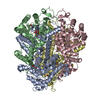
- Structure visualization
| Structure viewer | Molecule: MolmilJmol/JSmol |
|---|
- Downloads & links
-Download
| PDBx/mmCIF format | 3e5b.cif.gz | 304 KB | Display | PDBx/mmCIF format |
|---|---|---|---|---|
| PDB format | pdb3e5b.ent.gz | 246.6 KB | Display | PDB format |
| PDBx/mmJSON format | 3e5b.json.gz | Tree view | PDBx/mmJSON format | |
| Others |  Other downloads Other downloads |
-Validation report
| Summary document | 3e5b_validation.pdf.gz | 459.7 KB | Display | wwPDB validaton report |
|---|---|---|---|---|
| Full document | 3e5b_full_validation.pdf.gz | 480 KB | Display | |
| Data in XML | 3e5b_validation.xml.gz | 53.9 KB | Display | |
| Data in CIF | 3e5b_validation.cif.gz | 74.1 KB | Display | |
| Arichive directory | https://data.pdbj.org/pub/pdb/validation_reports/e5/3e5bftp://data.pdbj.org/pub/pdb/validation_reports/e5/3e5b | HTTPS FTP |
-Related structure data
| Similar structure data | |
|---|---|
| Other databases |


-Links
 PDBj
PDBj
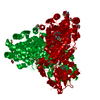
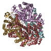
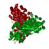
- Assembly
| Deposited unit | 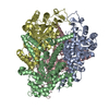
| ||||||||
|---|---|---|---|---|---|---|---|---|---|
| 1 |
| ||||||||
| Unit cell |
| ||||||||
| Details | authors state that the biological unit is the same as asymmetric unit. |
-Components
| #1: Protein | Mass: 47148.051 Da / Num. of mol.: 4 Source method: isolated from a genetically manipulated source Source: (gene. exp.) Brucella abortus (bacteria) / Strain: BIOVAR ABORTUS 2308 / Gene: aceA, BruAb1_1601 / Plasmid: AVA0421 / Production host: #2: Water | ChemComp-HOH / |  Mass: 18.015 Da / Num. of mol.: 102 / Source method: isolated from a natural source / Formula: H2O Mass: 18.015 Da / Num. of mol.: 102 / Source method: isolated from a natural source / Formula: H2O |
|---|
-Experimental details
-Experiment
| Experiment | Method: X-RAY DIFFRACTION / Number of used crystals: 1 |
|---|
- Sample preparation
| Crystal | Density Matthews: 2.56 Å3/Da / Density % sol: 52.02 % |
|---|---|
| Crystal grow | Temperature: 289 K / Method: vapor diffusion / pH: 8.5 Details: 30% PEG 4000, 0.1M TRIS pH 8.5, 0.2M LITHIUM SULFATE, VAPOR DIFFUSION, temperature 289K |
-Data collection
| Diffraction | Mean temperature: 100 K | ||||||||||||||||||||||||||||||||||||||||||||||||||||||||||||||||||
|---|---|---|---|---|---|---|---|---|---|---|---|---|---|---|---|---|---|---|---|---|---|---|---|---|---|---|---|---|---|---|---|---|---|---|---|---|---|---|---|---|---|---|---|---|---|---|---|---|---|---|---|---|---|---|---|---|---|---|---|---|---|---|---|---|---|---|---|
| Diffraction source | Source: SYNCHROTRON / Site: APS  / Beamline: 24-ID-C / Wavelength: 1 Å / Beamline: 24-ID-C / Wavelength: 1 Å | ||||||||||||||||||||||||||||||||||||||||||||||||||||||||||||||||||
| Detector | Type: ADSC QUANTUM 315 / Detector: CCD / Date: Jul 9, 2008 / Details: ADJUSTABLE FOCUSING MIRRORS | ||||||||||||||||||||||||||||||||||||||||||||||||||||||||||||||||||
| Radiation | Monochromator: Double Crystal Monochromator / Protocol: SINGLE WAVELENGTH / Scattering type: x-ray | ||||||||||||||||||||||||||||||||||||||||||||||||||||||||||||||||||
| Radiation wavelength | Wavelength: 1 Å / Relative weight: 1 | ||||||||||||||||||||||||||||||||||||||||||||||||||||||||||||||||||
| Reflection | Resolution: 2.37→50 Å / Num. obs: 77975 / % possible obs: 99.6 % / Redundancy: 7.1 % / Rmerge(I) obs: 0.119 / Χ2: 0.955 | ||||||||||||||||||||||||||||||||||||||||||||||||||||||||||||||||||
| Reflection shell |
|
-Phasing
| Phasing | Method: molecular replacement | |||||||||
|---|---|---|---|---|---|---|---|---|---|---|
| Phasing MR | Rfactor: 39.49 / Model details: Phaser MODE: MR_AUTO
|
- Processing
| Software |
| ||||||||||||||||||||||||||||||||||||||||||||||||||||||||||||||||||||||||||||||||||||||||||
|---|---|---|---|---|---|---|---|---|---|---|---|---|---|---|---|---|---|---|---|---|---|---|---|---|---|---|---|---|---|---|---|---|---|---|---|---|---|---|---|---|---|---|---|---|---|---|---|---|---|---|---|---|---|---|---|---|---|---|---|---|---|---|---|---|---|---|---|---|---|---|---|---|---|---|---|---|---|---|---|---|---|---|---|---|---|---|---|---|---|---|---|
| Refinement | Method to determine structure: MOLECULAR REPLACEMENT / Resolution: 2.37→20 Å / Cor.coef. Fo:Fc: 0.953 / Cor.coef. Fo:Fc free: 0.934 / WRfactor Rfree: 0.252 / WRfactor Rwork: 0.208 / Occupancy max: 1 / Occupancy min: 0.5 / FOM work R set: 0.812 / SU B: 8.614 / SU ML: 0.2 / SU R Cruickshank DPI: 0.358 / SU Rfree: 0.249 / Cross valid method: THROUGHOUT / σ(F): 0 / ESU R: 0.358 / ESU R Free: 0.249 / Stereochemistry target values: MAXIMUM LIKELIHOOD / Details: HYDROGENS HAVE BEEN ADDED IN THE RIDING POSITIONS
| ||||||||||||||||||||||||||||||||||||||||||||||||||||||||||||||||||||||||||||||||||||||||||
| Solvent computation | Ion probe radii: 0.8 Å / Shrinkage radii: 0.8 Å / VDW probe radii: 1.2 Å / Solvent model: MASK | ||||||||||||||||||||||||||||||||||||||||||||||||||||||||||||||||||||||||||||||||||||||||||
| Displacement parameters | Biso max: 87.94 Å2 / Biso mean: 57.361 Å2 / Biso min: 35.06 Å2
| ||||||||||||||||||||||||||||||||||||||||||||||||||||||||||||||||||||||||||||||||||||||||||
| Refinement step | Cycle: LAST / Resolution: 2.37→20 Å
| ||||||||||||||||||||||||||||||||||||||||||||||||||||||||||||||||||||||||||||||||||||||||||
| Refine LS restraints |
| ||||||||||||||||||||||||||||||||||||||||||||||||||||||||||||||||||||||||||||||||||||||||||
| LS refinement shell | Resolution: 2.371→2.432 Å / Total num. of bins used: 20
|
