 Movie
Movie Controller

[English] 日本語
 Yorodumi
- PDB-3e23: Crystal structure of the RPA2492 protein in complex with SAM from... -
+ Open data

- Basic information
| Entry | Database: PDB / ID: 3.0E+23 | ||||||
|---|---|---|---|---|---|---|---|
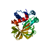
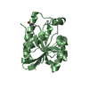
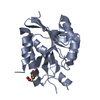
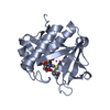
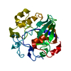
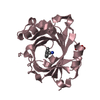
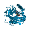
| Title | Crystal structure of the RPA2492 protein in complex with SAM from Rhodopseudomonas palustris, Northeast Structural Genomics Consortium Target RpR299 | ||||||
 Components Components | uncharacterized protein RPA2492 | ||||||
 Keywords Keywords | structural genomics / unknown function / alpha-beta protein / PSI-2 / Protein Structure Initiative / Northeast Structural Genomics Consortium / NESG | ||||||
| Function / homology |  Function and homology information Function and homology information | ||||||
| Biological species |  Rhodopseudomonas palustris (phototrophic) Rhodopseudomonas palustris (phototrophic) | ||||||
| Method |  X-RAY DIFFRACTION / SYNCHROTRON / SAD / Resolution: 1.6 Å X-RAY DIFFRACTION / SYNCHROTRON / SAD / Resolution: 1.6 Å | ||||||
 Authors Authors | Forouhar, F. / Chen, Y. / Seetharaman, J. / Mao, L. / Xiao, R. / Foote, E.L. / Ciccosanti, C. / Wang, H. / Tong, S. / Everett, J.K. ...Forouhar, F. / Chen, Y. / Seetharaman, J. / Mao, L. / Xiao, R. / Foote, E.L. / Ciccosanti, C. / Wang, H. / Tong, S. / Everett, J.K. / Acton, T.B. / Montelione, G.T. / Tong, L. / Hunt, J.F. / Northeast Structural Genomics Consortium (NESG) | ||||||
 Citation Citation | Journal: To be Published Title: Crystal structure of the RPA2492 protein in complex with SAM from Rhodopseudomonas palustris, Northeast Structural Genomics Consortium Target RpR299 Authors: Forouhar, F. / Chen, Y. / Seetharaman, J. / Mao, L. / Xiao, R. / Foote, E.L. / Ciccosanti, C. / Wang, H. / Tong, S. / K Everett, J. / Acton, T.B. / Montelione, G.T. / Tong, L. / Hunt, J.F. | ||||||
| History |
|
- Structure visualization
| Structure viewer | Molecule: MolmilJmol/JSmol |
|---|
- Downloads & links
-Download
| PDBx/mmCIF format | 3e23.cif.gz | 62.5 KB | Display | PDBx/mmCIF format |
|---|---|---|---|---|
| PDB format | pdb3e23.ent.gz | 43.8 KB | Display | PDB format |
| PDBx/mmJSON format | 3e23.json.gz | Tree view | PDBx/mmJSON format | |
| Others |  Other downloads Other downloads |
-Validation report
| Arichive directory | https://data.pdbj.org/pub/pdb/validation_reports/e2/3e23ftp://data.pdbj.org/pub/pdb/validation_reports/e2/3e23 | HTTPS FTP |
|---|
-Related structure data
| Similar structure data | |
|---|---|
| Other databases |


-Links
 PDBj
PDBj
- Assembly
| Deposited unit | 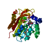
| ||||||||
|---|---|---|---|---|---|---|---|---|---|
| 1 |
| ||||||||
| Unit cell |
|
-Components
| #1: Protein | Mass: 23756.932 Da / Num. of mol.: 1 Source method: isolated from a genetically manipulated source Source: (gene. exp.) Rhodopseudomonas palustris (phototrophic)Strain: CGA009 / Gene: RPA2492 / Plasmid: pET21 / Production host: | ||||||
|---|---|---|---|---|---|---|---|
| #2: Chemical | ChemComp-SO4 /   Mass: 96.063 Da / Num. of mol.: 5 / Source method: obtained synthetically / Formula: SO4 Mass: 96.063 Da / Num. of mol.: 5 / Source method: obtained synthetically / Formula: SO4#3: Chemical | ChemComp-SAM / |   Mass: 398.437 Da / Num. of mol.: 1 / Source method: obtained synthetically / Formula: C15H22N6O5S Mass: 398.437 Da / Num. of mol.: 1 / Source method: obtained synthetically / Formula: C15H22N6O5S#4: Water | ChemComp-HOH / |  Mass: 18.015 Da / Num. of mol.: 317 / Source method: isolated from a natural source / Formula: H2O Mass: 18.015 Da / Num. of mol.: 317 / Source method: isolated from a natural source / Formula: H2OHas protein modification | Y | |
-Experimental details
-Experiment
| Experiment | Method: X-RAY DIFFRACTION / Number of used crystals: 1 |
|---|
- Sample preparation
| Crystal | Density Matthews: 2.45 Å3/Da / Density % sol: 49.85 % |
|---|---|
| Crystal grow | Temperature: 277 K / pH: 7.5 Details: Protein solution: 10 mM Tris (pH 7.5), 100 mM sodium chloride, 5 mM DTT, and 5 mM S-adenosyl-methionine. Resevoir solution: 100 mM HEPES (pH 7.5) and 2 M ammonium sulfate. , microbatch under ...Details: Protein solution: 10 mM Tris (pH 7.5), 100 mM sodium chloride, 5 mM DTT, and 5 mM S-adenosyl-methionine. Resevoir solution: 100 mM HEPES (pH 7.5) and 2 M ammonium sulfate. , microbatch under oil, temperature 277K |
-Data collection
| Diffraction | Mean temperature: 100 K |
|---|---|
| Diffraction source | Source: SYNCHROTRON / Site: NSLS  / Beamline: X4A / Wavelength: 0.97902 Å / Beamline: X4A / Wavelength: 0.97902 Å |
| Detector | Type: ADSC QUANTUM 4 / Detector: CCD / Date: Jul 28, 2008 / Details: mirrors |
| Radiation | Monochromator: Si 111 CHANNEL / Protocol: SINGLE WAVELENGTH / Monochromatic (M) / Laue (L): M / Scattering type: x-ray |
| Radiation wavelength | Wavelength: 0.97902 Å / Relative weight: 1 |
| Reflection | Resolution: 1.6→30 Å / Num. all: 60048 / Num. obs: 54164 / % possible obs: 90.2 % / Observed criterion σ(F): 2 / Observed criterion σ(I): 2 / Redundancy: 4.1 % / Biso Wilson estimate: 9.9 Å2 / Rmerge(I) obs: 0.049 / Rsym value: 0.057 / Net I/σ(I): 33.94 |
| Reflection shell | Resolution: 1.6→1.66 Å / Redundancy: 2.4 % / Rmerge(I) obs: 0.074 / Mean I/σ(I) obs: 12.77 / Num. unique all: 6000 / Rsym value: 0.109 / % possible all: 66.3 |
- Processing
| Software |
| ||||||||||||||||||||||||||||||||||||||||||||||||||||||||||||
|---|---|---|---|---|---|---|---|---|---|---|---|---|---|---|---|---|---|---|---|---|---|---|---|---|---|---|---|---|---|---|---|---|---|---|---|---|---|---|---|---|---|---|---|---|---|---|---|---|---|---|---|---|---|---|---|---|---|---|---|---|---|
| Refinement | Method to determine structure: SAD / Resolution: 1.6→19.86 Å / Rfactor Rfree error: 0.003 / Data cutoff high absF: 465336.69 / Data cutoff low absF: 2 / Isotropic thermal model: RESTRAINED / Cross valid method: THROUGHOUT / σ(F): 2 / σ(I): 2 / Stereochemistry target values: Engh & Huber
| ||||||||||||||||||||||||||||||||||||||||||||||||||||||||||||
| Solvent computation | Solvent model: FLAT MODEL / Bsol: 45.9086 Å2 / ksol: 0.4 e/Å3 | ||||||||||||||||||||||||||||||||||||||||||||||||||||||||||||
| Displacement parameters | Biso mean: 13 Å2
| ||||||||||||||||||||||||||||||||||||||||||||||||||||||||||||
| Refine analyze |
| ||||||||||||||||||||||||||||||||||||||||||||||||||||||||||||
| Refinement step | Cycle: LAST / Resolution: 1.6→19.86 Å
| ||||||||||||||||||||||||||||||||||||||||||||||||||||||||||||
| Refine LS restraints |
| ||||||||||||||||||||||||||||||||||||||||||||||||||||||||||||
| LS refinement shell | Resolution: 1.6→1.66 Å / Rfactor Rfree error: 0.01 / Total num. of bins used: 10
|
