 Movie
Movie Controller

[English] 日本語
 Yorodumi
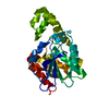
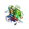
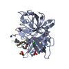
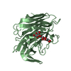
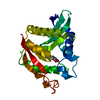
- PDB-3e1x: The Crystal Structure of Apo Prostasin at 1.7 Angstroms Resolution -
+ Open data

- Basic information
| Entry | Database: PDB / ID: 3e1x | ||||||
|---|---|---|---|---|---|---|---|
| Title | The Crystal Structure of Apo Prostasin at 1.7 Angstroms Resolution | ||||||
 Components Components | Prostasin | ||||||
 Keywords Keywords | HYDROLASE / Prostasin / ENaC / HCAP-1 / Channel Activating Protease / Cell membrane / Glycoprotein / Membrane / Protease / Secreted / Serine protease / Transmembrane / Zymogen | ||||||
| Function / homology |  Function and homology information Function and homology informationFormation of the cornified envelope / positive regulation of sodium ion transport / Hydrolases; Acting on peptide bonds (peptidases); Serine endopeptidases / serine-type peptidase activity / serine-type endopeptidase activity / proteolysis / extracellular space / extracellular exosome / extracellular region / plasma membrane Similarity search - Function | ||||||
| Biological species |  Homo sapiens (human) Homo sapiens (human) | ||||||
| Method |  X-RAY DIFFRACTION / SYNCHROTRON / MOLECULAR REPLACEMENT / Resolution: 1.7 Å X-RAY DIFFRACTION / SYNCHROTRON / MOLECULAR REPLACEMENT / Resolution: 1.7 Å | ||||||
 Authors Authors | Spraggon, G. / Hornsby, M. / Shipway, A. / Harris, J.L. / Lesley, S.A. | ||||||
 Citation Citation | Journal: Protein Sci. / Year: 2009 Title: Active site conformational changes of prostasin provide a new mechanism of protease regulation by divalent cations. Authors: Spraggon, G. / Hornsby, M. / Shipway, A. / Tully, D.C. / Bursulaya, B. / Danahay, H. / Harris, J.L. / Lesley, S.A. | ||||||
| History |
|
- Structure visualization
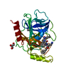
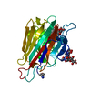
| Structure viewer | Molecule: MolmilJmol/JSmol |
|---|
- Downloads & links
-Download
| PDBx/mmCIF format | 3e1x.cif.gz | 65.8 KB | Display | PDBx/mmCIF format |
|---|---|---|---|---|
| PDB format | pdb3e1x.ent.gz | 47.2 KB | Display | PDB format |
| PDBx/mmJSON format | 3e1x.json.gz | Tree view | PDBx/mmJSON format | |
| Others |  Other downloads Other downloads |
-Validation report
| Summary document | 3e1x_validation.pdf.gz | 426.4 KB | Display | wwPDB validaton report |
|---|---|---|---|---|
| Full document | 3e1x_full_validation.pdf.gz | 428.3 KB | Display | |
| Data in XML | 3e1x_validation.xml.gz | 13.5 KB | Display | |
| Data in CIF | 3e1x_validation.cif.gz | 19.5 KB | Display | |
| Arichive directory | https://data.pdbj.org/pub/pdb/validation_reports/e1/3e1xftp://data.pdbj.org/pub/pdb/validation_reports/e1/3e1x | HTTPS FTP |
-Related structure data
| Related structure data | 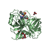 3e0nC  3fvfC  3gylC  3gymC 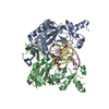 1eonS S: Starting model for refinement C: citing same article ( |
|---|---|
| Similar structure data |


-Links
 PDBj
PDBj- Assembly
| Deposited unit | 
| ||||||||
|---|---|---|---|---|---|---|---|---|---|
| 1 |
| ||||||||
| Unit cell |
|
-Components
| #1: Protein | Mass: 29608.002 Da / Num. of mol.: 1 / Fragment: UNP residues 45-305 Source method: isolated from a genetically manipulated source Source: (gene. exp.) Homo sapiens (human) / Gene: PRSS8 / Production host:   Spodoptera frugiperda (fall armyworm) Spodoptera frugiperda (fall armyworm)References: UniProt: Q16651, Hydrolases; Acting on peptide bonds (peptidases); Serine endopeptidases | ||||
|---|---|---|---|---|---|
| #2: Chemical |   Mass: 92.094 Da / Num. of mol.: 2 / Source method: obtained synthetically / Formula: C3H8O3 Mass: 92.094 Da / Num. of mol.: 2 / Source method: obtained synthetically / Formula: C3H8O3#3: Water | ChemComp-HOH / |  Mass: 18.015 Da / Num. of mol.: 199 / Source method: isolated from a natural source / Formula: H2O Mass: 18.015 Da / Num. of mol.: 199 / Source method: isolated from a natural source / Formula: H2OHas protein modification | Y | |
-Experimental details
-Experiment
| Experiment | Method: X-RAY DIFFRACTION / Number of used crystals: 1 |
|---|
- Sample preparation
| Crystal | Density Matthews: 2.02 Å3/Da / Density % sol: 39.16 % |
|---|---|
| Crystal grow | Temperature: 277 K / Method: vapor diffusion, sitting drop / pH: 6 Details: 30% Peg-6000 in 0.1M MES buffer, pH 6.0, VAPOR DIFFUSION, SITTING DROP, temperature 277K |
-Data collection
| Diffraction | Mean temperature: 100 K |
|---|---|
| Diffraction source | Source: SYNCHROTRON / Site: ALS  / Beamline: 5.0.3 / Wavelength: 1 Å / Beamline: 5.0.3 / Wavelength: 1 Å |
| Detector | Type: ADSC QUANTUM 210 / Detector: CCD / Date: Jun 28, 2006 |
| Radiation | Monochromator: graphite / Protocol: SINGLE WAVELENGTH / Monochromatic (M) / Laue (L): M / Scattering type: x-ray |
| Radiation wavelength | Wavelength: 1 Å / Relative weight: 1 |
| Reflection | Resolution: 1.7→50 Å / Num. obs: 26345 / % possible obs: 96.4 % / Observed criterion σ(F): 0 / Observed criterion σ(I): 0 / Redundancy: 3.5 % / Rmerge(I) obs: 0.059 / Rsym value: 0.059 / Net I/σ(I): 11.67 |
| Reflection shell | Resolution: 1.7→1.76 Å / Redundancy: 2.7 % / Rmerge(I) obs: 0.627 / Mean I/σ(I) obs: 1.08 / Num. unique all: 2047 / Rsym value: 0.627 / % possible all: 76.3 |
- Processing
| Software |
| |||||||||||||||||||||||||
|---|---|---|---|---|---|---|---|---|---|---|---|---|---|---|---|---|---|---|---|---|---|---|---|---|---|---|
| Refinement | Method to determine structure: MOLECULAR REPLACEMENT Starting model: 1eon Resolution: 1.7→45.819 Å / Isotropic thermal model: isotropic / Cross valid method: THROUGHOUT / σ(F): 0 / σ(I): 0 / Stereochemistry target values: Engh & Huber
| |||||||||||||||||||||||||
| Displacement parameters | Biso mean: 12.86 Å2
| |||||||||||||||||||||||||
| Refinement step | Cycle: LAST / Resolution: 1.7→45.819 Å
| |||||||||||||||||||||||||
| LS refinement shell | Resolution: 1.7→1.7 Å
|
