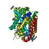
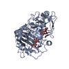
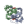
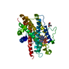
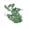
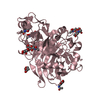
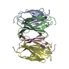
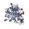
ジャーナル: Proc Natl Acad Sci U S A / 年: 2008 タイトル: Structural basis for induced formation of the inflammatory mediator prostaglandin E2. 著者: Caroline Jegerschöld / Sven-Christian Pawelzik / Pasi Purhonen / Priyaranjan Bhakat / Karina Roxana Gheorghe / Nobuhiko Gyobu / Kaoru Mitsuoka / Ralf Morgenstern / Per-Johan Jakobsson / Hans Hebert / 要旨: Prostaglandins (PG) are bioactive lipids produced from arachidonic acid via the action of cyclooxygenases and terminal PG synthases. Microsomal prostaglandin E synthase 1 (MPGES1) constitutes an ...Prostaglandins (PG) are bioactive lipids produced from arachidonic acid via the action of cyclooxygenases and terminal PG synthases. Microsomal prostaglandin E synthase 1 (MPGES1) constitutes an inducible glutathione-dependent integral membrane protein that catalyzes the oxidoreduction of cyclooxygenase derived PGH(2) into PGE(2). MPGES1 has been implicated in a number of human diseases or pathological conditions, such as rheumatoid arthritis, fever, and pain, and is therefore regarded as a primary target for development of novel antiinflammatory drugs. To provide a structural basis for insight in the catalytic mechanism, we determined the structure of MPGES1 in complex with glutathione by electron crystallography from 2D crystals induced in the presence of phospholipids. Together with results from site-directed mutagenesis and activity measurements, we can thereby demonstrate the role of specific amino acid residues. Glutathione is found to bind in a U-shaped conformation at the interface between subunits in the protein trimer. It is exposed to a site facing the lipid bilayer, which forms the specific environment for the oxidoreduction of PGH(2) to PGE(2) after displacement of the cytoplasmic half of the N-terminal transmembrane helix. Hence, insight into the dynamic behavior of MPGES1 and homologous membrane proteins in inflammation and detoxification is provided.
 ムービー
ムービー コントローラー


 データを開く
 基本情報
基本情報 要素
要素 キーワード
キーワード 機能・相同性情報
 Homo sapiens (ヒト)
 データ登録者
 引用
引用
 構造の表示
構造の表示 ダウンロードとリンク
 その他のダウンロード
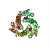
 PDBj
PDBj


 集合体
集合体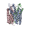


 分子量: 307.323 Da / 分子数: 3 / 由来タイプ: 合成 / 式: C10H17N3O6S
 試料調製
試料調製 FIELD EMISSION GUN / 加速電圧: 200 kV / 照射モード: FLOOD BEAM
 解析
解析