[English] 日本語
 Yorodumi
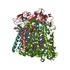
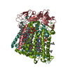
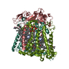
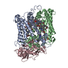
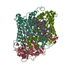
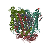
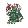
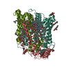
- PDB-3duq: E(L212)A, D(L213)A, N(M5)D triple mutant structure of photosynthe... -
+ Open data
- Basic information
| Entry | Database: PDB / ID: 3duq | ||||||
|---|---|---|---|---|---|---|---|
| Title | E(L212)A, D(L213)A, N(M5)D triple mutant structure of photosynthetic reaction center from Rhodobacter sphaeroides | ||||||
 Components Components | (Reaction center protein ...) x 3 | ||||||
 Keywords Keywords | PHOTOSYNTHESIS / Mutant photosynthetic reaction center / Phenotypic revertant / Proton transfer / membrane protein | ||||||
| Function / homology |  Function and homology information Function and homology informationplasma membrane-derived chromatophore membrane / plasma membrane light-harvesting complex / bacteriochlorophyll binding / photosynthetic electron transport in photosystem II / : / photosynthesis, light reaction / metal ion binding Similarity search - Function | ||||||
| Biological species |  Rhodobacter sphaeroides (bacteria) Rhodobacter sphaeroides (bacteria) | ||||||
| Method |  X-RAY DIFFRACTION / X-RAY DIFFRACTION /  SYNCHROTRON / SYNCHROTRON /  MOLECULAR REPLACEMENT / Resolution: 2.7 Å MOLECULAR REPLACEMENT / Resolution: 2.7 Å | ||||||
 Authors Authors | Pokkuluri, P.R. / Schiffer, M. | ||||||
 Citation Citation |  Journal: To be Published Journal: To be PublishedTitle: Structural description of compensatory mutations that restore proton transfer pathways to the L212A-L213A mutant bacterial reaction center Authors: Pokkuluri, P.R. / Laible, P.D. / Ginell, S.L. / Hanson, D.K. / Schiffer, M. | ||||||
| History |
|
- Structure visualization
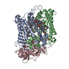
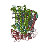
| Structure viewer | Molecule:  Molmil Molmil Jmol/JSmol Jmol/JSmol |
|---|
- Downloads & links
- Download
| PDBx/mmCIF format |  3duq.cif.gz 3duq.cif.gz | 204.9 KB | Display |  PDBx/mmCIF format PDBx/mmCIF format |
|---|---|---|---|---|
| PDB format |  pdb3duq.ent.gz pdb3duq.ent.gz | 157.7 KB | Display |  PDB format PDB format |
| PDBx/mmJSON format |  3duq.json.gz 3duq.json.gz | Tree view |  PDBx/mmJSON format PDBx/mmJSON format | |
| Others |  Other downloads Other downloads |
-Validation report
| Arichive directory |  https://data.pdbj.org/pub/pdb/validation_reports/du/3duq https://data.pdbj.org/pub/pdb/validation_reports/du/3duq ftp://data.pdbj.org/pub/pdb/validation_reports/du/3duq ftp://data.pdbj.org/pub/pdb/validation_reports/du/3duq | HTTPS FTP |
|---|
-Related structure data
| Related structure data | 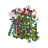 1k6nS S: Starting model for refinement |
|---|---|
| Similar structure data |
- Links
Links
- Assembly
| Deposited unit | 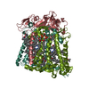
| ||||||||
|---|---|---|---|---|---|---|---|---|---|
| 1 |
| ||||||||
| Unit cell |
| ||||||||
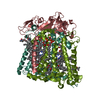
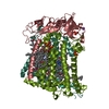
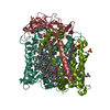
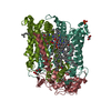
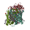
| Details | Authors state that the photosynthetic reaction center is a complex made up of three protein chains (L,M,H) and several co-factors. |
- Components
-Reaction center protein ... , 3 types, 3 molecules LMH
| #1: Protein | Mass: 31244.346 Da / Num. of mol.: 1 / Mutation: E(L212)A, D(L213)A, N(M5)D Source method: isolated from a genetically manipulated source Source: (gene. exp.)  Rhodobacter sphaeroides (bacteria) / Gene: pufL / Production host: Rhodobacter sphaeroides (bacteria) / Gene: pufL / Production host:  Rhodobacter sphaeroides (bacteria) / References: UniProt: P0C0Y8 Rhodobacter sphaeroides (bacteria) / References: UniProt: P0C0Y8 |
|---|---|
| #2: Protein | Mass: 35366.543 Da / Num. of mol.: 1 Source method: isolated from a genetically manipulated source Source: (gene. exp.)  Rhodobacter sphaeroides (bacteria) / Gene: pufM / Production host: Rhodobacter sphaeroides (bacteria) / Gene: pufM / Production host:  Rhodobacter sphaeroides (bacteria) / References: UniProt: P0C0Y9 Rhodobacter sphaeroides (bacteria) / References: UniProt: P0C0Y9 |
| #3: Protein | Mass: 28066.322 Da / Num. of mol.: 1 Source method: isolated from a genetically manipulated source Source: (gene. exp.)  Rhodobacter sphaeroides (bacteria) / Gene: puhA / Production host: Rhodobacter sphaeroides (bacteria) / Gene: puhA / Production host:  Rhodobacter sphaeroides (bacteria) / References: UniProt: P0C0Y7 Rhodobacter sphaeroides (bacteria) / References: UniProt: P0C0Y7 |
-Non-polymers , 8 types, 170 molecules 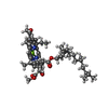
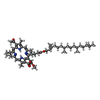
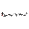
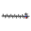

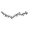


| #4: Chemical | ChemComp-BCL / #5: Chemical | #6: Chemical | #7: Chemical | ChemComp-LDA / #8: Chemical | ChemComp-FE / | #9: Chemical | ChemComp-SPN / | #10: Chemical | ChemComp-CDL / | #11: Water | ChemComp-HOH / | |
|---|
-Experimental details
-Experiment
| Experiment | Method:  X-RAY DIFFRACTION / Number of used crystals: 4 X-RAY DIFFRACTION / Number of used crystals: 4 |
|---|
- Sample preparation
| Crystal | Density % sol: 78.44 % |
|---|---|
| Crystal grow | Temperature: 293 K / Method: vapor diffusion, sitting drop / pH: 7.5 Details: Potassium phosphate, LDAO, Heptane triol, Dioxane, pH 7.5, VAPOR DIFFUSION, SITTING DROP, temperature 293K |
-Data collection
| Diffraction | Mean temperature: 273 K |
|---|---|
| Diffraction source | Source:  SYNCHROTRON / Site: SYNCHROTRON / Site:  APS APS  / Beamline: 19-ID / Beamline: 19-ID |
| Detector | Type: CUSTOM-MADE / Detector: CCD / Date: Apr 9, 2002 |
| Radiation | Protocol: SINGLE WAVELENGTH / Monochromatic (M) / Laue (L): M / Scattering type: x-ray |
| Radiation wavelength | Relative weight: 1 |
| Reflection | Resolution: 2.7→30 Å / Num. obs: 55881 / % possible obs: 97 % / Observed criterion σ(I): -3 / Redundancy: 4.7 % / Rmerge(I) obs: 0.092 / Net I/σ(I): 23 |
| Reflection shell | Resolution: 2.7→2.8 Å / Redundancy: 2 % / Rmerge(I) obs: 0.391 / Mean I/σ(I) obs: 2.6 / Num. unique all: 3899 / % possible all: 77 |
- Processing
| Software |
| |||||||||||||||||||||||||
|---|---|---|---|---|---|---|---|---|---|---|---|---|---|---|---|---|---|---|---|---|---|---|---|---|---|---|
| Refinement | Method to determine structure:  MOLECULAR REPLACEMENT MOLECULAR REPLACEMENTStarting model: 1k6n with M5Asn side chain truncated at CB Resolution: 2.7→30 Å / σ(F): 0 / Stereochemistry target values: Engh & Huber Details: After refitting, only isotropic B-factor refinement was done with CNS. No positional refinement was done
| |||||||||||||||||||||||||
| Refinement step | Cycle: LAST / Resolution: 2.7→30 Å
|
 Movie
Movie Controller







 PDBj
PDBj


