 Movie
Movie Controller

[English] 日本語
 Yorodumi
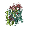
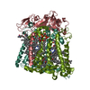
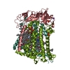
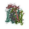
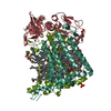
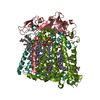
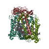
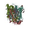
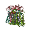
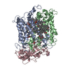
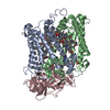
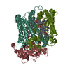
- PDB-1pss: CRYSTALLOGRAPHIC ANALYSES OF SITE-DIRECTED MUTANTS OF THE PHOTOSY... -
+ Open data

- Basic information
| Entry | Database: PDB / ID: 1pss | ||||||
|---|---|---|---|---|---|---|---|
| Title | CRYSTALLOGRAPHIC ANALYSES OF SITE-DIRECTED MUTANTS OF THE PHOTOSYNTHETIC REACTION CENTER FROM RHODOBACTER SPHAEROIDES | ||||||
 Components Components | (PHOTOSYNTHETIC REACTION ...) x 3 | ||||||
 Keywords Keywords | PHOTOSYNTHETIC REACTION CENTER | ||||||
| Function / homology |  Function and homology information Function and homology information: / : / plasma membrane-derived chromatophore membrane / plasma membrane light-harvesting complex / bacteriochlorophyll binding / photosynthetic electron transport in photosystem II / photosynthesis, light reaction / membrane => GO:0016020 / metal ion binding Similarity search - Function | ||||||
| Biological species |  Rhodobacter sphaeroides (bacteria) Rhodobacter sphaeroides (bacteria) | ||||||
| Method |  X-RAY DIFFRACTION / Resolution: 3 Å X-RAY DIFFRACTION / Resolution: 3 Å | ||||||
 Authors Authors | Chirino, A.J. / Feher, G. / Rees, D.C. | ||||||
 Citation Citation | Journal: Biochemistry / Year: 1994 Title: Crystallographic analyses of site-directed mutants of the photosynthetic reaction center from Rhodobacter sphaeroides. Authors: Chirino, A.J. / Lous, E.J. / Huber, M. / Allen, J.P. / Schenck, C.C. / Paddock, M.L. / Feher, G. / Rees, D.C. #1: Journal: Proc.Natl.Acad.Sci.USA / Year: 1988Title: Structure of the Reaction Center from Rhodobacter Sphaeroides R-26 and 2.4.1:Protein-Cofactor (Bacteriochlorophyll,Bacteriopheophytin, and Carotenoid) Interactions Authors: Yeates, T.O. / Komiya, H. / Chirino, A. / Rees, D.C. / Allen, J.P. / Feher, G. #2: Journal: Annu.Rev.Biochem. / Year: 1989Title: The Bacterial Photosynthetic Reaction Center as a Model for Membrane Proteins Authors: Rees, D.C. / Komiya, H. / Yeates, T.O. / Allen, J.P. / Feher, G. #3: Journal: Nature / Year: 1989Title: Structure and Function of Bacterial Photosynthetic Reaction Centers Authors: Feher, G. / Allen, J.P. / Okamura, M.Y. / Rees, D.C. #4: Journal: Proc.Natl.Acad.Sci.USA / Year: 1988Title: Structure of the Reaction Center from Rhodobacter Sphaeroides R-26 and 2.4.1: Symmetry Relations and Sequence Comparisons between Different Species Authors: Komiya, H. / Yeates, T.O. / Rees, D.C. / Allen, J.P. / Feher, G. #5: Journal: Proc.Natl.Acad.Sci.USA / Year: 1988Title: Structure of the Reaction Center from Rhodobacter Sphaeroides R-26: Protein-Cofactor (Quinones and Fe2+) Interactions Authors: Allen, J.P. / Feher, G. / Yeates, T.O. / Komiya, H. / Rees, D.C. #6: Journal: Proc.Natl.Acad.Sci.USA / Year: 1987Title: Structure of the Reaction Center from Rhodobacter Sphaeroides R-26: Membrane-Protein Interactions Authors: Yeates, T.O. / Komiya, H. / Rees, D.C. / Allen, J.P. / Feher, G. #7: Journal: Proc.Natl.Acad.Sci.USA / Year: 1987Title: Structure of the Reaction Center from Rhodobacter Sphaeroides R-26: The Protein Subunits Authors: Allen, J.P. / Feher, G. / Yeates, T.O. / Komiya, H. / Rees, D.C. #8: Journal: Proc.Natl.Acad.Sci.USA / Year: 1987Title: Structure of the Reaction Center from Rhodobacter Sphaeroides R-26: The Cofactors Authors: Allen, J.P. / Feher, G. / Yeates, T.O. / Komiya, H. / Rees, D.C. #9: Journal: Proc.Natl.Acad.Sci.USA / Year: 1986Title: Structure Homology of the Reaction Center from Rhodopseudomonas Sphaeroides and Rhodopseudomonas Viridis as Determined by X-Ray Diffraction Authors: Allen, J.P. / Feher, G. / Yeates, T.O. / Rees, D.C. / Desenhofer, J. / Michel, H. / Huber, R. | ||||||
| History |
|
- Structure visualization
| Structure viewer | Molecule: MolmilJmol/JSmol |
|---|
- Downloads & links
-Download
| PDBx/mmCIF format | 1pss.cif.gz | 187.9 KB | Display | PDBx/mmCIF format |
|---|---|---|---|---|
| PDB format | pdb1pss.ent.gz | 145.4 KB | Display | PDB format |
| PDBx/mmJSON format | 1pss.json.gz | Tree view | PDBx/mmJSON format | |
| Others |  Other downloads Other downloads |
-Validation report
| Arichive directory | https://data.pdbj.org/pub/pdb/validation_reports/ps/1pssftp://data.pdbj.org/pub/pdb/validation_reports/ps/1pss | HTTPS FTP |
|---|
-Related structure data


-Links
 PDBj
PDBj


- Assembly
| Deposited unit | 
| ||||||||
|---|---|---|---|---|---|---|---|---|---|
| 1 |
| ||||||||
| Unit cell |
| ||||||||
| Atom site foot note | 1: LEU L 269 - PRO L 270 OMEGA = 34.32 PEPTIDE BOND DEVIATES SIGNIFICANTLY FROM TRANS CONFORMATION 2: CIS PROLINE - PRO M 49 3: PRO M 96 - PRO M 97 OMEGA =125.41 PEPTIDE BOND DEVIATES SIGNIFICANTLY FROM TRANS CONFORMATION 4: CIS PROLINE - PRO M 99 5: ARG H 83 - PRO H 84 OMEGA =251.89 PEPTIDE BOND DEVIATES SIGNIFICANTLY FROM TRANS CONFORMATION 6: ALA H 245 - PRO H 246 OMEGA =149.81 PEPTIDE BOND DEVIATES SIGNIFICANTLY FROM TRANS CONFORMATION |
-Components
-PHOTOSYNTHETIC REACTION ... , 3 types, 3 molecules LMH
| #1: Protein | Mass: 29795.633 Da / Num. of mol.: 1 Source method: isolated from a genetically manipulated source Source: (gene. exp.) Rhodobacter sphaeroides (bacteria) / References: UniProt: P02954, UniProt: P0C0Y8*PLUS |
|---|---|
| #2: Protein | Mass: 33209.246 Da / Num. of mol.: 1 Source method: isolated from a genetically manipulated source Source: (gene. exp.) Rhodobacter sphaeroides (bacteria) / References: UniProt: P02953, UniProt: P0C0Y9*PLUS |
| #3: Protein | Mass: 25691.557 Da / Num. of mol.: 1 Source method: isolated from a genetically manipulated source Source: (gene. exp.) Rhodobacter sphaeroides (bacteria) / References: UniProt: P11846, UniProt: P0C0Y7*PLUS |
-Non-polymers , 5 types, 10 molecules 


| #4: Chemical | ChemComp-BCL /  Mass: 911.504 Da / Num. of mol.: 4 / Source method: obtained synthetically / Formula: C55H74MgN4O6 Mass: 911.504 Da / Num. of mol.: 4 / Source method: obtained synthetically / Formula: C55H74MgN4O6#5: Chemical |  Mass: 889.215 Da / Num. of mol.: 2 / Source method: obtained synthetically / Formula: C55H76N4O6 Mass: 889.215 Da / Num. of mol.: 2 / Source method: obtained synthetically / Formula: C55H76N4O6#6: Chemical |  Mass: 863.343 Da / Num. of mol.: 2 / Source method: obtained synthetically / Formula: C59H90O4 Mass: 863.343 Da / Num. of mol.: 2 / Source method: obtained synthetically / Formula: C59H90O4#7: Chemical | ChemComp-FE / |  Mass: 55.845 Da / Num. of mol.: 1 / Source method: obtained synthetically / Formula: Fe Mass: 55.845 Da / Num. of mol.: 1 / Source method: obtained synthetically / Formula: Fe#8: Chemical | ChemComp-CRT / |  Mass: 596.925 Da / Num. of mol.: 1 / Source method: obtained synthetically / Formula: C42H60O2 Mass: 596.925 Da / Num. of mol.: 1 / Source method: obtained synthetically / Formula: C42H60O2 |
|---|
-Experimental details
-Experiment
| Experiment | Method: X-RAY DIFFRACTION |
|---|
- Sample preparation
| Crystal | Density Matthews: 4.27 Å3/Da / Density % sol: 71.21 % | ||||||||||||||||||||||||||||||||||||||||||||||||||||||||||||||||||||||||||||||||||||
|---|---|---|---|---|---|---|---|---|---|---|---|---|---|---|---|---|---|---|---|---|---|---|---|---|---|---|---|---|---|---|---|---|---|---|---|---|---|---|---|---|---|---|---|---|---|---|---|---|---|---|---|---|---|---|---|---|---|---|---|---|---|---|---|---|---|---|---|---|---|---|---|---|---|---|---|---|---|---|---|---|---|---|---|---|---|
| Crystal grow | *PLUS pH: 8 / Method: vapor diffusion, hanging dropDetails: taken from Allen, J.P. et al (1986). Proc. Natl. Acad. Sci., 83, 8589-8593. | ||||||||||||||||||||||||||||||||||||||||||||||||||||||||||||||||||||||||||||||||||||
| Components of the solutions | *PLUS
|
-Data collection
| Radiation | Scattering type: x-ray |
|---|---|
| Radiation wavelength | Relative weight: 1 |
| Reflection | *PLUS Highest resolution: 3 Å / Num. obs: 21518 / % possible obs: 68.9 % / Num. measured all: 61853 / Rmerge(I) obs: 0.077 |
- Processing
| Software |
| ||||||||||||||||||||||||||||||||||||||||||||||||||||||||||||
|---|---|---|---|---|---|---|---|---|---|---|---|---|---|---|---|---|---|---|---|---|---|---|---|---|---|---|---|---|---|---|---|---|---|---|---|---|---|---|---|---|---|---|---|---|---|---|---|---|---|---|---|---|---|---|---|---|---|---|---|---|---|
| Refinement | Rfactor Rwork: 0.223 / Rfactor obs: 0.223 / Highest resolution: 3 Å Details: THE STRUCTURE OF THE H-CHAIN IS BASED MAINLY ON MOLECULAR REPLACEMENT FROM THE RPS. VIRIDIS REACTION CENTER STRUCTURE AND IS LESS RELIABLE THAN REST OF THE STRUCTURE. IN GENERAL THE ...Details: THE STRUCTURE OF THE H-CHAIN IS BASED MAINLY ON MOLECULAR REPLACEMENT FROM THE RPS. VIRIDIS REACTION CENTER STRUCTURE AND IS LESS RELIABLE THAN REST OF THE STRUCTURE. IN GENERAL THE TRANSMEMBRANE REGION OF THE STRUCTURE IS MORE RELIABLE THAN THE HYDROPHILIC REGIONS OF THE MOLECULE. | ||||||||||||||||||||||||||||||||||||||||||||||||||||||||||||
| Refinement step | Cycle: LAST / Highest resolution: 3 Å
| ||||||||||||||||||||||||||||||||||||||||||||||||||||||||||||
| Refine LS restraints |
| ||||||||||||||||||||||||||||||||||||||||||||||||||||||||||||
| Software | *PLUS Name: X-PLOR / Classification: refinement | ||||||||||||||||||||||||||||||||||||||||||||||||||||||||||||
| Refinement | *PLUS Highest resolution: 3 Å / Num. reflection obs: 18066 / σ(F): 2 / Rfactor obs: 0.223 | ||||||||||||||||||||||||||||||||||||||||||||||||||||||||||||
| Solvent computation | *PLUS | ||||||||||||||||||||||||||||||||||||||||||||||||||||||||||||
| Displacement parameters | *PLUS | ||||||||||||||||||||||||||||||||||||||||||||||||||||||||||||
| Refine LS restraints | *PLUS Type: x_angle_d / Dev ideal: 3.41 |
