 Movie
Movie Controller


+ Open data

- Basic information
| Entry | Database: PDB / ID: 1m3x | ||||||
|---|---|---|---|---|---|---|---|
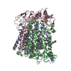
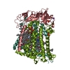
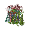
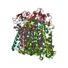
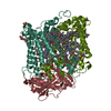
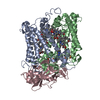
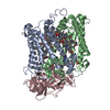
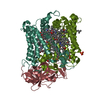
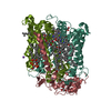
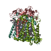
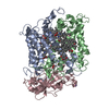
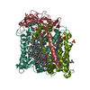
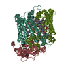
| Title | Photosynthetic Reaction Center From Rhodobacter Sphaeroides | ||||||
 Components Components | (Photosynthetic Reaction center protein ...) x 3 | ||||||
 Keywords Keywords | PHOTOSYNTHESIS / alpha helix / membrane protein | ||||||
| Function / homology |  Function and homology information Function and homology information: / : / plasma membrane-derived chromatophore membrane / plasma membrane light-harvesting complex / bacteriochlorophyll binding / photosynthetic electron transport in photosystem II / photosynthesis, light reaction / membrane => GO:0016020 / metal ion binding Similarity search - Function | ||||||
| Biological species |  Rhodobacter sphaeroides (bacteria) Rhodobacter sphaeroides (bacteria) | ||||||
| Method |  X-RAY DIFFRACTION / SYNCHROTRON / MOLECULAR REPLACEMENT / Resolution: 2.55 Å X-RAY DIFFRACTION / SYNCHROTRON / MOLECULAR REPLACEMENT / Resolution: 2.55 Å | ||||||
 Authors Authors | Camara-Artigas, A. / Brune, D. / Allen, J.P. | ||||||
 Citation Citation | Journal: Proc.Natl.Acad.Sci.USA / Year: 2002 Title: Interactions between lipids and bacterial reaction centers determined by protein crystallography. Authors: Camara-Artigas, A. / Brune, D. / Allen, J.P. #1: Journal: Acta Crystallogr.,Sect.D / Year: 2001Title: Individual interactions influence the crystalline order for membrane proteins Authors: Camara-Artigas, A. / Magee, C.L. / Williams, J.C. / Allen, J.P. | ||||||
| History |
| ||||||
| Remark 600 | HETEROGEN THE FOLLOWING LIGANDS HAVE ATOMS MISSING DUE TO LACK OF ELECTRON DENSITY: BCL 850, U10 ...HETEROGEN THE FOLLOWING LIGANDS HAVE ATOMS MISSING DUE TO LACK OF ELECTRON DENSITY: BCL 850, U10 857, U10 858, CDL 900, PC2 901, GGD 902. |
- Structure visualization
| Structure viewer | Molecule: MolmilJmol/JSmol |
|---|
- Downloads & links
-Download
| PDBx/mmCIF format | 1m3x.cif.gz | 206.7 KB | Display | PDBx/mmCIF format |
|---|---|---|---|---|
| PDB format | pdb1m3x.ent.gz | 156.8 KB | Display | PDB format |
| PDBx/mmJSON format | 1m3x.json.gz | Tree view | PDBx/mmJSON format | |
| Others |  Other downloads Other downloads |
-Validation report
| Arichive directory | https://data.pdbj.org/pub/pdb/validation_reports/m3/1m3xftp://data.pdbj.org/pub/pdb/validation_reports/m3/1m3x | HTTPS FTP |
|---|
-Related structure data
| Related structure data | 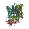 1qovS S: Starting model for refinement |
|---|---|
| Similar structure data |


-Links
 PDBj
PDBj


- Assembly
| Deposited unit | 
| ||||||||
|---|---|---|---|---|---|---|---|---|---|
| 1 |
| ||||||||
| Unit cell |
|
-Components
-Photosynthetic Reaction center protein ... , 3 types, 3 molecules LMH
| #1: Protein | Mass: 31346.389 Da / Num. of mol.: 1 Source method: isolated from a genetically manipulated source Source: (gene. exp.) Rhodobacter sphaeroides (bacteria) / Production host: Rhodobacter sphaeroides (bacteria) / References: UniProt: P02954, UniProt: P0C0Y8*PLUS |
|---|---|
| #2: Protein | Mass: 34398.543 Da / Num. of mol.: 1 Source method: isolated from a genetically manipulated source Source: (gene. exp.) Rhodobacter sphaeroides (bacteria) / Production host: Rhodobacter sphaeroides (bacteria) / References: UniProt: P02953, UniProt: P0C0Y9*PLUS |
| #3: Protein | Mass: 28066.322 Da / Num. of mol.: 1 Source method: isolated from a genetically manipulated source Source: (gene. exp.) Rhodobacter sphaeroides (bacteria) / Production host: Rhodobacter sphaeroides (bacteria) / References: UniProt: P11846, UniProt: P0C0Y7*PLUS |
-Non-polymers , 10 types, 217 molecules 


| #4: Chemical | ChemComp-BCL /  Mass: 911.504 Da / Num. of mol.: 4 / Source method: obtained synthetically / Formula: C55H74MgN4O6 Mass: 911.504 Da / Num. of mol.: 4 / Source method: obtained synthetically / Formula: C55H74MgN4O6#5: Chemical |  Mass: 889.215 Da / Num. of mol.: 2 / Source method: obtained synthetically / Formula: C55H76N4O6 Mass: 889.215 Da / Num. of mol.: 2 / Source method: obtained synthetically / Formula: C55H76N4O6#6: Chemical |  Mass: 863.343 Da / Num. of mol.: 2 / Source method: obtained synthetically / Formula: C59H90O4 Mass: 863.343 Da / Num. of mol.: 2 / Source method: obtained synthetically / Formula: C59H90O4#7: Chemical | ChemComp-PC1 / |  Mass: 790.145 Da / Num. of mol.: 1 / Source method: obtained synthetically / Formula: C44H88NO8P / Comment: phospholipid*YM Mass: 790.145 Da / Num. of mol.: 1 / Source method: obtained synthetically / Formula: C44H88NO8P / Comment: phospholipid*YM#8: Chemical | ChemComp-FE / |  Mass: 55.845 Da / Num. of mol.: 1 / Source method: obtained synthetically / Formula: Fe Mass: 55.845 Da / Num. of mol.: 1 / Source method: obtained synthetically / Formula: Fe#9: Chemical | ChemComp-CL / |  Mass: 35.453 Da / Num. of mol.: 1 / Source method: obtained synthetically / Formula: Cl Mass: 35.453 Da / Num. of mol.: 1 / Source method: obtained synthetically / Formula: Cl#10: Chemical | ChemComp-SPO / |  Mass: 568.914 Da / Num. of mol.: 1 / Source method: obtained synthetically / Formula: C41H60O Mass: 568.914 Da / Num. of mol.: 1 / Source method: obtained synthetically / Formula: C41H60O#11: Chemical | ChemComp-CDL / |  Mass: 1464.043 Da / Num. of mol.: 1 / Source method: obtained synthetically / Formula: C81H156O17P2 / Comment: phospholipid*YM Mass: 1464.043 Da / Num. of mol.: 1 / Source method: obtained synthetically / Formula: C81H156O17P2 / Comment: phospholipid*YM#12: Chemical | ChemComp-GGD / |  Mass: 959.294 Da / Num. of mol.: 1 / Source method: obtained synthetically / Formula: C52H94O15 Mass: 959.294 Da / Num. of mol.: 1 / Source method: obtained synthetically / Formula: C52H94O15#13: Water | ChemComp-HOH / | Mass: 18.015 Da / Num. of mol.: 203 / Source method: isolated from a natural source / Formula: H2O |
|---|
-Experimental details
-Experiment
| Experiment | Method: X-RAY DIFFRACTION / Number of used crystals: 1 |
|---|
- Sample preparation
| Crystal grow | Temperature: 298 K / Method: vapor diffusion, sitting drop / pH: 7.4 Details: POTASIUM PHOSPHATE, LDAO, HEPTANE TRIOL, pH 7.4, VAPOR DIFFUSION, SITTING DROP, temperature 298K | ||||||||||||||||||||||||||||||||||||||||||
|---|---|---|---|---|---|---|---|---|---|---|---|---|---|---|---|---|---|---|---|---|---|---|---|---|---|---|---|---|---|---|---|---|---|---|---|---|---|---|---|---|---|---|---|
| Crystal grow | *PLUS | ||||||||||||||||||||||||||||||||||||||||||
| Components of the solutions | *PLUS
|
-Data collection
| Diffraction | Mean temperature: 298 K |
|---|---|
| Diffraction source | Source: SYNCHROTRON / Site: SSRL  / Beamline: BL7-1 / Wavelength: 1.08 Å / Beamline: BL7-1 / Wavelength: 1.08 Å |
| Detector | Type: MARRESEARCH / Detector: IMAGE PLATE / Date: Jan 1, 2000 Details: 58 cm long, Pt-coated, fused silica, vertical focus mirror; Cyclindrically bent triangular Si(111) asymmetric cut |
| Radiation | Monochromator: horizontal focus monochromator / Protocol: SINGLE WAVELENGTH / Monochromatic (M) / Laue (L): M / Scattering type: x-ray |
| Radiation wavelength | Wavelength: 1.08 Å / Relative weight: 1 |
| Reflection | Resolution: 2.55→30.83 Å / Num. obs: 68970 / % possible obs: 97.2 % / Observed criterion σ(F): 0 / Observed criterion σ(I): 0 / Redundancy: 4.2 % / Biso Wilson estimate: 23.8 Å2 / Rmerge(I) obs: 0.126 / Rsym value: 0.109 / Net I/σ(I): 4.3 |
| Reflection shell | Resolution: 2.55→2.69 Å / Redundancy: 3.6 % / Rmerge(I) obs: 0.406 / Mean I/σ(I) obs: 1.5 / Num. unique all: 34173 / Rsym value: 0.364 / % possible all: 92.5 |
| Reflection | *PLUS Lowest resolution: 30 Å / Num. obs: 69691 / Num. measured all: 564825 |
| Reflection shell | *PLUS Lowest resolution: 2.65 Å / % possible obs: 92.5 % |
- Processing
| Software |
| ||||||||||||||||||||||||||||||||||||||||||||||||||||||||||||
|---|---|---|---|---|---|---|---|---|---|---|---|---|---|---|---|---|---|---|---|---|---|---|---|---|---|---|---|---|---|---|---|---|---|---|---|---|---|---|---|---|---|---|---|---|---|---|---|---|---|---|---|---|---|---|---|---|---|---|---|---|---|
| Refinement | Method to determine structure: MOLECULAR REPLACEMENT Starting model: pdb entry 1QOV Resolution: 2.55→29.91 Å / Rfactor Rfree error: 0.003 / Isotropic thermal model: RESTRAINED / Cross valid method: THROUGHOUT / σ(F): 2 / σ(I): 0 / Stereochemistry target values: Engh & Huber
| ||||||||||||||||||||||||||||||||||||||||||||||||||||||||||||
| Solvent computation | Solvent model: FLAT MODEL / Bsol: 61.9782 Å2 / ksol: 0.326599 e/Å3 | ||||||||||||||||||||||||||||||||||||||||||||||||||||||||||||
| Displacement parameters | Biso mean: 36.3 Å2
| ||||||||||||||||||||||||||||||||||||||||||||||||||||||||||||
| Refine analyze | Luzzati coordinate error free: 0.29 Å / Luzzati sigma a free: 0.3 Å | ||||||||||||||||||||||||||||||||||||||||||||||||||||||||||||
| Refinement step | Cycle: LAST / Resolution: 2.55→29.91 Å
| ||||||||||||||||||||||||||||||||||||||||||||||||||||||||||||
| Refine LS restraints |
| ||||||||||||||||||||||||||||||||||||||||||||||||||||||||||||
| LS refinement shell | Resolution: 2.55→2.71 Å / Rfactor Rfree error: 0.008 / Total num. of bins used: 6
| ||||||||||||||||||||||||||||||||||||||||||||||||||||||||||||
| Xplor file |
| ||||||||||||||||||||||||||||||||||||||||||||||||||||||||||||
| Refinement | *PLUS Lowest resolution: 30 Å / Rfactor Rfree: 0.205 / Rfactor Rwork: 0.183 | ||||||||||||||||||||||||||||||||||||||||||||||||||||||||||||
| Solvent computation | *PLUS | ||||||||||||||||||||||||||||||||||||||||||||||||||||||||||||
| Displacement parameters | *PLUS | ||||||||||||||||||||||||||||||||||||||||||||||||||||||||||||
| Refine LS restraints | *PLUS
|
