 Movie
Movie Controller

[English] 日本語
 Yorodumi
- PDB-3dsy: E(L212)Q mutant structure of photosynthetic reaction center from ... -
+ Open data

- Basic information
| Entry | Database: PDB / ID: 3dsy | ||||||
|---|---|---|---|---|---|---|---|
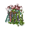
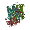
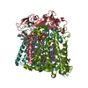
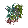
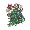
| Title | E(L212)Q mutant structure of photosynthetic reaction center from Rhodobacter sphaeroides | ||||||
 Components Components | (Reaction center protein ...) x 3 | ||||||
 Keywords Keywords | PHOTOSYNTHESIS / Mutant Photosynthetic reaction center / Proton transfer / membrane protein | ||||||
| Function / homology |  Function and homology information Function and homology informationplasma membrane-derived chromatophore membrane / plasma membrane light-harvesting complex / bacteriochlorophyll binding / photosynthetic electron transport in photosystem II / photosynthesis, light reaction / metal ion binding Similarity search - Function | ||||||
| Biological species |  Rhodobacter sphaeroides (bacteria) Rhodobacter sphaeroides (bacteria) | ||||||
| Method |  X-RAY DIFFRACTION / MOLECULAR REPLACEMENT / Resolution: 3 Å X-RAY DIFFRACTION / MOLECULAR REPLACEMENT / Resolution: 3 Å | ||||||
 Authors Authors | Pokkuluri, P.R. / Schiffer, M. | ||||||
 Citation Citation | Journal: To be Published Title: Restoring Proton transfer in L212Q mutant of photosynthetic reaction center Authors: Pokkuluri, P.R. / Laible, P.D. / Hanson, D.K. / Schiffer, M. | ||||||
| History |
|
- Structure visualization
| Structure viewer | Molecule: MolmilJmol/JSmol |
|---|
- Downloads & links
-Download
| PDBx/mmCIF format | 3dsy.cif.gz | 204.2 KB | Display | PDBx/mmCIF format |
|---|---|---|---|---|
| PDB format | pdb3dsy.ent.gz | 157.1 KB | Display | PDB format |
| PDBx/mmJSON format | 3dsy.json.gz | Tree view | PDBx/mmJSON format | |
| Others |  Other downloads Other downloads |
-Validation report
| Arichive directory | https://data.pdbj.org/pub/pdb/validation_reports/ds/3dsyftp://data.pdbj.org/pub/pdb/validation_reports/ds/3dsy | HTTPS FTP |
|---|
-Related structure data
| Related structure data |  1k6lS S: Starting model for refinement |
|---|---|
| Similar structure data |


-Links
 PDBj
PDBj


- Assembly
| Deposited unit | 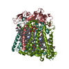
| ||||||||
|---|---|---|---|---|---|---|---|---|---|
| 1 |
| ||||||||
| Unit cell |
| ||||||||
| Details | Authors state that the photosynthetic reaction center is a complex made up of three protein chains (L,M,H) and several co-factors. |
-Components
-Reaction center protein ... , 3 types, 3 molecules LMH
| #1: Protein | Mass: 31345.404 Da / Num. of mol.: 1 / Mutation: EL212Q Source method: isolated from a genetically manipulated source Source: (gene. exp.) Rhodobacter sphaeroides (bacteria) / Gene: pufL / Production host: Rhodobacter sphaeroides (bacteria) / References: UniProt: P0C0Y8 |
|---|---|
| #2: Protein | Mass: 35365.559 Da / Num. of mol.: 1 Source method: isolated from a genetically manipulated source Source: (gene. exp.) Rhodobacter sphaeroides (bacteria) / Gene: pufM / Production host: Rhodobacter sphaeroides (bacteria) / References: UniProt: P0C0Y9 |
| #3: Protein | Mass: 28066.322 Da / Num. of mol.: 1 Source method: isolated from a genetically manipulated source Source: (gene. exp.) Rhodobacter sphaeroides (bacteria) / Gene: puhA / Production host: Rhodobacter sphaeroides (bacteria) / References: UniProt: P0C0Y7 |
-Non-polymers , 8 types, 154 molecules 


| #4: Chemical | ChemComp-BCL /  Mass: 911.504 Da / Num. of mol.: 4 / Source method: obtained synthetically / Formula: C55H74MgN4O6 Mass: 911.504 Da / Num. of mol.: 4 / Source method: obtained synthetically / Formula: C55H74MgN4O6#5: Chemical |  Mass: 889.215 Da / Num. of mol.: 2 / Source method: obtained synthetically / Formula: C55H76N4O6 Mass: 889.215 Da / Num. of mol.: 2 / Source method: obtained synthetically / Formula: C55H76N4O6#6: Chemical |  Mass: 863.343 Da / Num. of mol.: 2 / Source method: obtained synthetically / Formula: C59H90O4 Mass: 863.343 Da / Num. of mol.: 2 / Source method: obtained synthetically / Formula: C59H90O4#7: Chemical | ChemComp-LDA /  Mass: 229.402 Da / Num. of mol.: 4 / Source method: obtained synthetically / Formula: C14H31NO / Comment: LDAO, detergent*YM Mass: 229.402 Da / Num. of mol.: 4 / Source method: obtained synthetically / Formula: C14H31NO / Comment: LDAO, detergent*YM#8: Chemical | ChemComp-FE / |  Mass: 55.845 Da / Num. of mol.: 1 / Source method: obtained synthetically / Formula: Fe Mass: 55.845 Da / Num. of mol.: 1 / Source method: obtained synthetically / Formula: Fe#9: Chemical | ChemComp-SPN / |  Mass: 594.993 Da / Num. of mol.: 1 / Source method: obtained synthetically / Formula: C41H70O2 Mass: 594.993 Da / Num. of mol.: 1 / Source method: obtained synthetically / Formula: C41H70O2#10: Chemical | ChemComp-CDL / |  Mass: 1464.043 Da / Num. of mol.: 1 / Source method: obtained synthetically / Formula: C81H156O17P2 / Comment: phospholipid*YM Mass: 1464.043 Da / Num. of mol.: 1 / Source method: obtained synthetically / Formula: C81H156O17P2 / Comment: phospholipid*YM#11: Water | ChemComp-HOH / | Mass: 18.015 Da / Num. of mol.: 139 / Source method: isolated from a natural source / Formula: H2O |
|---|
-Experimental details
-Experiment
| Experiment | Method: X-RAY DIFFRACTION / Number of used crystals: 8 |
|---|
- Sample preparation
| Crystal | Density Matthews: 5.79 Å3/Da / Density % sol: 78.75 % |
|---|---|
| Crystal grow | Temperature: 293 K / Method: vapor diffusion, sitting drop / pH: 7.5 Details: Potassium phosphate, LDAO, Heptane triol, Dioxane, pH 7.5, VAPOR DIFFUSION, SITTING DROP, temperature 293K |
-Data collection
| Diffraction | Mean temperature: 293 K |
|---|---|
| Diffraction source | Source: ROTATING ANODE / Type: RIGAKU RU200 / Wavelength: 1.54 Å |
| Detector | Type: RIGAKU RAXIS IIC / Detector: IMAGE PLATE / Date: May 6, 2005 / Details: mirrors |
| Radiation | Protocol: SINGLE WAVELENGTH / Monochromatic (M) / Laue (L): M / Scattering type: x-ray |
| Radiation wavelength | Wavelength: 1.54 Å / Relative weight: 1 |
| Reflection | Resolution: 3→30 Å / Num. obs: 42457 / % possible obs: 98 % / Observed criterion σ(I): -3 / Redundancy: 3.4 % / Rmerge(I) obs: 0.097 / Net I/σ(I): 13.7 |
| Reflection shell | Resolution: 3→3.11 Å / Redundancy: 2.6 % / Rmerge(I) obs: 0.292 / Mean I/σ(I) obs: 3.9 / Num. unique all: 3706 / % possible all: 93 |
- Processing
| Software |
| ||||||||||||||||
|---|---|---|---|---|---|---|---|---|---|---|---|---|---|---|---|---|---|
| Refinement | Method to determine structure: MOLECULAR REPLACEMENT Starting model: PDB entry 1k6l Resolution: 3→30 Å / σ(F): 0 / Stereochemistry target values: Engh & Huber Details: 1K6L was used as starting model for rigid-body refinement. After refitting the coordinates only B-factor refinement was done with CNS. No positional refinement was done
| ||||||||||||||||
| Refinement step | Cycle: LAST / Resolution: 3→30 Å
|
