 Movie
Movie Controller

[English] 日本語
 Yorodumi
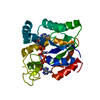
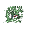
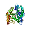
- PDB-3dt6: Crystal Structure of Bovin Brain Platelet Activating Factor Acety... -
+ Open data

- Basic information
| Entry | Database: PDB / ID: 3dt6 | ||||||
|---|---|---|---|---|---|---|---|
| Title | Crystal Structure of Bovin Brain Platelet Activating Factor Acetylhydrolase Covalently Inhibited by Paraoxon | ||||||
 Components Components | Brain Platelet-activating factor acetylhydrolase IB subunit alpha | ||||||
 Keywords Keywords | HYDROLASE / PLATELET ACTIVATING FACTOR ACETYLHYDROLASE / PAF-AH IB / ALPHA-1 SUBUNIT / LIS1 / GROUP VIII PHOSPHOLIPASE A2 / 26 kDa / PARAOXON / Cytoplasm / Lipid degradation / PLATELET FACTOR | ||||||
| Function / homology |  Function and homology information Function and homology informationplatelet-activating factor acetyltransferase activity / 1-alkyl-2-acetylglycerophosphocholine esterase / 1-alkyl-2-acetylglycerophosphocholine esterase complex / COPI-independent Golgi-to-ER retrograde traffic / 1-alkyl-2-acetylglycerophosphocholine esterase activity / lipid catabolic process / spermatogenesis / protein heterodimerization activity / protein homodimerization activity / cytoplasm Similarity search - Function | ||||||
| Biological species |  | ||||||
| Method |  X-RAY DIFFRACTION / MOLECULAR REPLACEMENT / Resolution: 2.1 Å X-RAY DIFFRACTION / MOLECULAR REPLACEMENT / Resolution: 2.1 Å | ||||||
 Authors Authors | Epstein, T.M. / Samanta, U. / Bahnson, B.J. | ||||||
 Citation Citation | Journal: Biochemistry / Year: 2009 Title: Crystal structures of brain group-VIII phospholipase A2 in nonaged complexes with the organophosphorus nerve agents soman and sarin. Authors: Epstein, T.M. / Samanta, U. / Kirby, S.D. / Cerasoli, D.M. / Bahnson, B.J. #1: Journal: Nature / Year: 1997Title: Brain Acetylhydrolase that Inactivates Platelet-Activating Factor is a G-Protein-Like Trimer Authors: Ho, Y.S. / Swenson, L. / Derewenda, U. / Serre, L. / Wei, Y. / Dauter, Z. / Hattori, M. / Adachi, T. / Aoki, J. / Arai, H. / Inoue, K. / Derewenda, Z.S. | ||||||
| History |
|
- Structure visualization
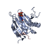
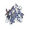
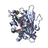
| Structure viewer | Molecule: MolmilJmol/JSmol |
|---|
- Downloads & links
-Download
| PDBx/mmCIF format | 3dt6.cif.gz | 56 KB | Display | PDBx/mmCIF format |
|---|---|---|---|---|
| PDB format | pdb3dt6.ent.gz | 39.7 KB | Display | PDB format |
| PDBx/mmJSON format | 3dt6.json.gz | Tree view | PDBx/mmJSON format | |
| Others |  Other downloads Other downloads |
-Validation report
| Arichive directory | https://data.pdbj.org/pub/pdb/validation_reports/dt/3dt6ftp://data.pdbj.org/pub/pdb/validation_reports/dt/3dt6 | HTTPS FTP |
|---|
-Related structure data
| Related structure data |  3dt8C 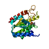 3dt9C  1wabS S: Starting model for refinement C: citing same article ( |
|---|---|
| Similar structure data |

-Links
 PDBj
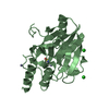
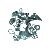
PDBj- Assembly
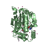
| Deposited unit | 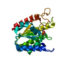
| ||||||||
|---|---|---|---|---|---|---|---|---|---|
| 1 |
| ||||||||
| Unit cell |
|
-Components
| #1: Protein | Mass: 25887.297 Da / Num. of mol.: 1 / Mutation: C55S Source method: isolated from a genetically manipulated source Source: (gene. exp.)  References: UniProt: Q29460, 1-alkyl-2-acetylglycerophosphocholine esterase |
|---|---|
| #2: Chemical | ChemComp-DEP /   Mass: 138.102 Da / Num. of mol.: 1 / Source method: obtained synthetically / Formula: C4H11O3P Mass: 138.102 Da / Num. of mol.: 1 / Source method: obtained synthetically / Formula: C4H11O3P |
| #3: Water | ChemComp-HOH /  Mass: 18.015 Da / Num. of mol.: 82 / Source method: isolated from a natural source / Formula: H2O Mass: 18.015 Da / Num. of mol.: 82 / Source method: isolated from a natural source / Formula: H2O |
| Has protein modification | Y |
-Experimental details
-Experiment
| Experiment | Method: X-RAY DIFFRACTION / Number of used crystals: 1 |
|---|
- Sample preparation
| Crystal | Density Matthews: 2.67 Å3/Da / Density % sol: 53.93 % |
|---|---|
| Crystal grow | Method: vapor diffusion, hanging drop / Details: VAPOR DIFFUSION, HANGING DROP |
-Data collection
| Diffraction | Mean temperature: 100 K |
|---|---|
| Diffraction source | Source: ROTATING ANODE / Type: RIGAKU RU300 / Wavelength: 1.54 |
| Detector | Type: RIGAKU / Detector: IMAGE PLATE / Date: Oct 24, 2003 / Details: OSMIC BLUE |
| Radiation | Protocol: SINGLE WAVELENGTH / Monochromatic (M) / Laue (L): M / Scattering type: x-ray |
| Radiation wavelength | Wavelength: 1.54 Å / Relative weight: 1 |
| Reflection | Resolution: 2.1→70.36 Å / Num. all: 15253 / Num. obs: 15253 / % possible obs: 92.1 % / Observed criterion σ(F): 1 / Observed criterion σ(I): 1 / Redundancy: 5.7 % / Rmerge(I) obs: 0.058 / Net I/σ(I): 28.28 |
| Reflection shell | Resolution: 2.1→2.18 Å / Redundancy: 5.2 % / Rmerge(I) obs: 0.354 / Mean I/σ(I) obs: 3.3 / % possible all: 90.6 |
- Processing
| Software |
| ||||||||||||||||||||||||||||||||||||||||||||||||||||||||||||||||||||||||||||||||||||||||||||||||||||||||||||||||||||||||||||||||||||||||||||||||||||||||||||||||||||||||||
|---|---|---|---|---|---|---|---|---|---|---|---|---|---|---|---|---|---|---|---|---|---|---|---|---|---|---|---|---|---|---|---|---|---|---|---|---|---|---|---|---|---|---|---|---|---|---|---|---|---|---|---|---|---|---|---|---|---|---|---|---|---|---|---|---|---|---|---|---|---|---|---|---|---|---|---|---|---|---|---|---|---|---|---|---|---|---|---|---|---|---|---|---|---|---|---|---|---|---|---|---|---|---|---|---|---|---|---|---|---|---|---|---|---|---|---|---|---|---|---|---|---|---|---|---|---|---|---|---|---|---|---|---|---|---|---|---|---|---|---|---|---|---|---|---|---|---|---|---|---|---|---|---|---|---|---|---|---|---|---|---|---|---|---|---|---|---|---|---|---|---|---|
| Refinement | Method to determine structure: MOLECULAR REPLACEMENT Starting model: PDB ENTRY 1WAB Resolution: 2.1→30 Å / Cor.coef. Fo:Fc: 0.945 / Cor.coef. Fo:Fc free: 0.947 / SU B: 3.251 / SU ML: 0.09 / Cross valid method: THROUGHOUT / σ(F): 1 / ESU R: 0.215 / ESU R Free: 0.159 / Stereochemistry target values: MAXIMUM LIKELIHOOD
| ||||||||||||||||||||||||||||||||||||||||||||||||||||||||||||||||||||||||||||||||||||||||||||||||||||||||||||||||||||||||||||||||||||||||||||||||||||||||||||||||||||||||||
| Solvent computation | Ion probe radii: 0.8 Å / Shrinkage radii: 0.8 Å / VDW probe radii: 1.4 Å / Solvent model: MASK | ||||||||||||||||||||||||||||||||||||||||||||||||||||||||||||||||||||||||||||||||||||||||||||||||||||||||||||||||||||||||||||||||||||||||||||||||||||||||||||||||||||||||||
| Displacement parameters | Biso mean: 30.629 Å2
| ||||||||||||||||||||||||||||||||||||||||||||||||||||||||||||||||||||||||||||||||||||||||||||||||||||||||||||||||||||||||||||||||||||||||||||||||||||||||||||||||||||||||||
| Refinement step | Cycle: LAST / Resolution: 2.1→30 Å
| ||||||||||||||||||||||||||||||||||||||||||||||||||||||||||||||||||||||||||||||||||||||||||||||||||||||||||||||||||||||||||||||||||||||||||||||||||||||||||||||||||||||||||
| Refine LS restraints |
| ||||||||||||||||||||||||||||||||||||||||||||||||||||||||||||||||||||||||||||||||||||||||||||||||||||||||||||||||||||||||||||||||||||||||||||||||||||||||||||||||||||||||||
| LS refinement shell | Resolution: 2.1→2.155 Å / Total num. of bins used: 20
|
