 Movie
Movie Controller

[English] 日本語
 Yorodumi
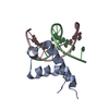
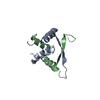
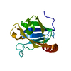
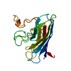
- PDB-3dlv: Structures of SRP54 and SRP19, the two proteins assembling the ri... -
+ Open data

- Basic information
| Entry | Database: PDB / ID: 3dlv | ||||||
|---|---|---|---|---|---|---|---|
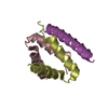
| Title | Structures of SRP54 and SRP19, the two proteins assembling the ribonucleic core of the Signal Recognition Particle from the archaeon Pyrococcus furiosus. | ||||||
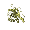
 Components Components | Signal recognition particle 19 kDa protein | ||||||
 Keywords Keywords | RNA BINDING PROTEIN / PROTEIN-RNA / SIGNAL RECOGNITION PARTICLE / Cytoplasm / Ribonucleoprotein / RNA-binding | ||||||
| Function / homology |  Function and homology information Function and homology informationsignal recognition particle / 7S RNA binding / SRP-dependent cotranslational protein targeting to membrane Similarity search - Function | ||||||
| Biological species |   Pyrococcus furiosus (archaea) Pyrococcus furiosus (archaea) | ||||||
| Method |  X-RAY DIFFRACTION / SYNCHROTRON / MOLECULAR REPLACEMENT / Resolution: 1.87 Å X-RAY DIFFRACTION / SYNCHROTRON / MOLECULAR REPLACEMENT / Resolution: 1.87 Å | ||||||
 Authors Authors | Egea, P.F. / Napetschnig, J. / Walter, P. / Stroud, R.M. | ||||||
 Citation Citation | Journal: Plos One / Year: 2008 Title: Structures of SRP54 and SRP19, the two proteins that organize the ribonucleic core of the signal recognition particle from Pyrococcus furiosus. Authors: Egea, P.F. / Napetschnig, J. / Walter, P. / Stroud, R.M. | ||||||
| History |
|
- Structure visualization
| Structure viewer | Molecule: MolmilJmol/JSmol |
|---|
- Downloads & links
-Download
| PDBx/mmCIF format | 3dlv.cif.gz | 52.3 KB | Display | PDBx/mmCIF format |
|---|---|---|---|---|
| PDB format | pdb3dlv.ent.gz | 37.4 KB | Display | PDB format |
| PDBx/mmJSON format | 3dlv.json.gz | Tree view | PDBx/mmJSON format | |
| Others |  Other downloads Other downloads |
-Validation report
| Summary document | 3dlv_validation.pdf.gz | 431.1 KB | Display | wwPDB validaton report |
|---|---|---|---|---|
| Full document | 3dlv_full_validation.pdf.gz | 432.5 KB | Display | |
| Data in XML | 3dlv_validation.xml.gz | 9.8 KB | Display | |
| Data in CIF | 3dlv_validation.cif.gz | 12.9 KB | Display | |
| Arichive directory | https://data.pdbj.org/pub/pdb/validation_reports/dl/3dlvftp://data.pdbj.org/pub/pdb/validation_reports/dl/3dlv | HTTPS FTP |
-Related structure data
| Related structure data |  3dluC  3dm5C  3dulS C: citing same article ( S: Starting model for refinement |
|---|---|
| Similar structure data |


-Links
 PDBj
PDBj- Assembly
| Deposited unit | 
| |||||||||
|---|---|---|---|---|---|---|---|---|---|---|
| 1 | 
| |||||||||
| 2 | 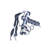
| |||||||||
| Unit cell |
| |||||||||
| Components on special symmetry positions |
|
-Components
| #1: Protein | Mass: 12291.387 Da / Num. of mol.: 2 Source method: isolated from a genetically manipulated source Source: (gene. exp.) Pyrococcus furiosus (archaea) / Strain: DSM3638 / Gene: srp19, PF1894 / Plasmid: pET29b / Production host:  #2: Water | ChemComp-HOH / |  Mass: 18.015 Da / Num. of mol.: 89 / Source method: isolated from a natural source / Formula: H2O Mass: 18.015 Da / Num. of mol.: 89 / Source method: isolated from a natural source / Formula: H2O |
|---|
-Experimental details
-Experiment
| Experiment | Method: X-RAY DIFFRACTION / Number of used crystals: 1 |
|---|
- Sample preparation
| Crystal | Density Matthews: 1.76 Å3/Da / Density % sol: 30.12 % |
|---|---|
| Crystal grow | Temperature: 293 K / Method: vapor diffusion, hanging drop / pH: 5 Details: 1.2-1.3M Na Malonate, 100 mM NaAcetate, pH 5.0, VAPOR DIFFUSION, HANGING DROP, temperature 293K |
-Data collection
| Diffraction | Mean temperature: 80 K |
|---|---|
| Diffraction source | Source: SYNCHROTRON / Site: ALS  / Beamline: 8.3.1 / Wavelength: 0.97949 Å / Beamline: 8.3.1 / Wavelength: 0.97949 Å |
| Detector | Type: ADSC QUANTUM 210 / Detector: CCD / Date: May 8, 2006 |
| Radiation | Protocol: SINGLE WAVELENGTH / Monochromatic (M) / Laue (L): M / Scattering type: x-ray |
| Radiation wavelength | Wavelength: 0.97949 Å / Relative weight: 1 |
| Reflection | Resolution: 1.87→50 Å / Num. all: 14745 / Num. obs: 14745 / % possible obs: 100 % / Observed criterion σ(F): 1 / Observed criterion σ(I): 1 / Redundancy: 7.9 % / Biso Wilson estimate: 15 Å2 / Rmerge(I) obs: 0.052 / Rsym value: 0.052 / Net I/σ(I): 22.3 |
| Reflection shell | Resolution: 1.87→1.94 Å / Redundancy: 6.2 % / Rmerge(I) obs: 0.513 / Mean I/σ(I) obs: 2 / Num. unique all: 1411 / Rsym value: 0.513 / % possible all: 98.3 |
- Processing
| Software |
| |||||||||||||||||||||||||||||||||||||||||||||||||
|---|---|---|---|---|---|---|---|---|---|---|---|---|---|---|---|---|---|---|---|---|---|---|---|---|---|---|---|---|---|---|---|---|---|---|---|---|---|---|---|---|---|---|---|---|---|---|---|---|---|---|
| Refinement | Method to determine structure: MOLECULAR REPLACEMENT Starting model: PDB ENTRY 3DUL Resolution: 1.87→47.8 Å / SU ML: 0.29 / Cross valid method: THROUGHOUT / σ(F): 0.85 / Phase error: 23.59 / Stereochemistry target values: MLHL
| |||||||||||||||||||||||||||||||||||||||||||||||||
| Solvent computation | Shrinkage radii: 0.9 Å / VDW probe radii: 1.11 Å / Solvent model: FLAT BULK SOLVENT MODEL / Bsol: 65.567 Å2 / ksol: 0.391 e/Å3 | |||||||||||||||||||||||||||||||||||||||||||||||||
| Displacement parameters | Biso mean: 22 Å2
| |||||||||||||||||||||||||||||||||||||||||||||||||
| Refinement step | Cycle: LAST / Resolution: 1.87→47.8 Å
| |||||||||||||||||||||||||||||||||||||||||||||||||
| Refine LS restraints |
| |||||||||||||||||||||||||||||||||||||||||||||||||
| LS refinement shell | Refine-ID: X-RAY DIFFRACTION / Total num. of bins used: 10
|
