 Movie
Movie Controller

[English] 日本語
 Yorodumi
- PDB-3dul: Crystal Structure Analysis of the O-methyltransferase from Bacill... -
+ Open data

- Basic information
| Entry | Database: PDB / ID: 3dul | ||||||
|---|---|---|---|---|---|---|---|
| Title | Crystal Structure Analysis of the O-methyltransferase from Bacillus cereus | ||||||
 Components Components | O-methyltransferase, putative | ||||||
 Keywords Keywords | TRANSFERASE / alternating of alpha and beta / Methyltransferase | ||||||
| Function / homology |  Function and homology information Function and homology informationS-adenosylmethionine-dependent methyltransferase activity / O-methyltransferase activity / methylation Similarity search - Function | ||||||
| Biological species |  | ||||||
| Method |  X-RAY DIFFRACTION / SYNCHROTRON / MAD / Resolution: 1.8 Å X-RAY DIFFRACTION / SYNCHROTRON / MAD / Resolution: 1.8 Å | ||||||
 Authors Authors | Cho, J.-H. / Rhee, S. | ||||||
 Citation Citation | Journal: J.Mol.Biol. / Year: 2008 Title: Structural and functional insights into O-methyltransferase from Bacillus cereus Authors: Cho, J.-H. / Park, Y. / Ahn, J.-H. / Lim, Y. / Rhee, S. | ||||||
| History |
|
- Structure visualization
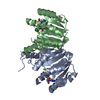
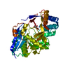
| Structure viewer | Molecule: MolmilJmol/JSmol |
|---|
- Downloads & links
-Download
| PDBx/mmCIF format | 3dul.cif.gz | 93.2 KB | Display | PDBx/mmCIF format |
|---|---|---|---|---|
| PDB format | pdb3dul.ent.gz | 72.2 KB | Display | PDB format |
| PDBx/mmJSON format | 3dul.json.gz | Tree view | PDBx/mmJSON format | |
| Others |  Other downloads Other downloads |
-Validation report
| Arichive directory | https://data.pdbj.org/pub/pdb/validation_reports/du/3dulftp://data.pdbj.org/pub/pdb/validation_reports/du/3dul | HTTPS FTP |
|---|
-Related structure data


-Links
 PDBj
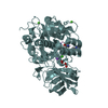
PDBj- Assembly
| Deposited unit | 
| ||||||||
|---|---|---|---|---|---|---|---|---|---|
| 1 |
| ||||||||
| Unit cell |
|
-Components
| #1: Protein | Mass: 24771.361 Da / Num. of mol.: 2 Source method: isolated from a genetically manipulated source Source: (gene. exp.) References: UniProt: Q739U3, Transferases; Transferring one-carbon groups; Methyltransferases #2: Water | ChemComp-HOH / |  Mass: 18.015 Da / Num. of mol.: 229 / Source method: isolated from a natural source / Formula: H2O Mass: 18.015 Da / Num. of mol.: 229 / Source method: isolated from a natural source / Formula: H2OHas protein modification | Y | |
|---|
-Experimental details
-Experiment
| Experiment | Method: X-RAY DIFFRACTION / Number of used crystals: 1 |
|---|
- Sample preparation
| Crystal | Density Matthews: 2.53 Å3/Da / Density % sol: 51.43 % |
|---|---|
| Crystal grow | Temperature: 295 K / Method: vapor diffusion, hanging drop / pH: 4.5 Details: 0.1M citric acid, 1.0M LiCl, 2% PEG 6000, 3% dioxane as an additive, pH 4.5, VAPOR DIFFUSION, HANGING DROP, temperature 295K |
-Data collection
| Diffraction | Mean temperature: 100 K | ||||||||||||
|---|---|---|---|---|---|---|---|---|---|---|---|---|---|
| Diffraction source | Source: SYNCHROTRON / Site: PAL/PLS  / Beamline: 4A / Wavelength: 0.97929, 0.97942, 0.97162 / Beamline: 4A / Wavelength: 0.97929, 0.97942, 0.97162 | ||||||||||||
| Detector | Type: ADSC QUANTUM 210 / Detector: CCD / Date: Dec 8, 2005 | ||||||||||||
| Radiation | Monochromator: Mirror / Protocol: MAD / Monochromatic (M) / Laue (L): M / Scattering type: x-ray | ||||||||||||
| Radiation wavelength |
| ||||||||||||
| Reflection | Resolution: 1.8→50 Å / Num. all: 45976 / Num. obs: 40986 / % possible obs: 99.9 % / Observed criterion σ(F): 2 / Redundancy: 7.2 % / Rmerge(I) obs: 0.072 / Net I/σ(I): 6 | ||||||||||||
| Reflection shell | Resolution: 1.8→1.86 Å / Redundancy: 7.2 % / Rmerge(I) obs: 0.359 / Mean I/σ(I) obs: 4.3 / Num. unique all: 4505 / % possible all: 100 |
- Processing
| Software |
| ||||||||||||||||||||
|---|---|---|---|---|---|---|---|---|---|---|---|---|---|---|---|---|---|---|---|---|---|
| Refinement | Method to determine structure: MAD / Resolution: 1.8→19.95 Å / Occupancy max: 1 / Occupancy min: 1 / Cross valid method: using a random selection of data / σ(F): 2
| ||||||||||||||||||||
| Solvent computation | Bsol: 47.367 Å2 | ||||||||||||||||||||
| Displacement parameters | Biso max: 50.98 Å2 / Biso mean: 26.86 Å2 / Biso min: 9.91 Å2
| ||||||||||||||||||||
| Refinement step | Cycle: LAST / Resolution: 1.8→19.95 Å
| ||||||||||||||||||||
| Refine LS restraints |
| ||||||||||||||||||||
| LS refinement shell | Resolution: 1.8→1.86 Å | ||||||||||||||||||||
| Xplor file |
|
