[English] 日本語
Yorodumi
- PDB-3di4: Crystal structure of a duf1989 family protein (spo0365) from sili... -

+
Open data


ID or keywords:

Loading...

-
Basic information

Entry
Database: PDB / ID: 3di4
TitleCrystal structure of a duf1989 family protein (spo0365) from silicibacter pomeroyi dss-3 at 1.60 A resolution
ComponentsUncharacterized Protein DUF1989
KeywordsMETAL BINDING PROTEIN / Structural genomics / Joint Center for Structural Genomics / JCSG / Protein Structure Initiative / PSI-2
Function / homologyDomain of unknown function DUF1989 / Domain of unknown function (DUF1989) / metal ion binding / DUF1989 domain-containing protein
Function and homology information
Biological speciesSilicibacter pomeroyi (bacteria)
MethodX-RAY DIFFRACTION / SYNCHROTRON / MAD / Resolution: 1.6 Å
AuthorsJoint Center for Structural Genomics (JCSG)
CitationJournal: To be published
Title: Crystal structure of protein of unknown function (DUF1989) (YP_165628.1) from SILICIBACTER POMEROYI DSS-3 at 1.60 A resolution
Authors: Joint Center for Structural Genomics (JCSG)
History
DepositionJun 19, 2008Deposition site: RCSB / Processing site: RCSB
Revision 1.0Jul 1, 2008Provider: repository / Type: Initial release
Revision 1.1Jul 13, 2011Group: Source and taxonomy / Version format compliance
Revision 1.2Oct 25, 2017Group: Refinement description / Category: software / Item: _software.classification / _software.name
Revision 1.3Jul 24, 2019Group: Data collection / Derived calculations / Refinement description
Category: software / struct_conn
Item: _software.classification / _software.contact_author ..._software.classification / _software.contact_author / _software.contact_author_email / _software.language / _software.location / _software.name / _software.type / _software.version / _struct_conn.pdbx_leaving_atom_flag
Revision 1.4Feb 1, 2023Group: Database references / Derived calculations
Category: database_2 / struct_conn ...database_2 / struct_conn / struct_ref_seq_dif / struct_site
Item: _database_2.pdbx_DOI / _database_2.pdbx_database_accession ..._database_2.pdbx_DOI / _database_2.pdbx_database_accession / _struct_conn.pdbx_dist_value / _struct_conn.ptnr1_auth_asym_id / _struct_conn.ptnr1_auth_comp_id / _struct_conn.ptnr1_auth_seq_id / _struct_conn.ptnr1_label_asym_id / _struct_conn.ptnr1_label_atom_id / _struct_conn.ptnr1_label_comp_id / _struct_conn.ptnr1_label_seq_id / _struct_conn.ptnr2_auth_asym_id / _struct_conn.ptnr2_auth_comp_id / _struct_conn.ptnr2_auth_seq_id / _struct_conn.ptnr2_label_asym_id / _struct_conn.ptnr2_label_atom_id / _struct_conn.ptnr2_label_comp_id / _struct_ref_seq_dif.details / _struct_site.pdbx_auth_asym_id / _struct_site.pdbx_auth_comp_id / _struct_site.pdbx_auth_seq_id

-
Structure visualization

Structure viewerMolecule:
MolmilJmol/JSmol

Downloads & links

-
Assembly

Deposited unit
A: Uncharacterized Protein DUF1989
B: Uncharacterized Protein DUF1989
hetero molecules


Theoretical massNumber of molelcules
Total (without water)63,65610
Polymers63,1952
Non-polymers4618
Water13,313739
1


  • Idetical with deposited unit
  • defined by author&software
TypeNameSymmetry operationNumber
identity operation1_555x,y,z1
Buried area9540 Å2
ΔGint-181 kcal/mol
Surface area19500 Å2
MethodPISA
Unit cell
Length a, b, c (Å)51.465, 95.764, 58.689
Angle α, β, γ (deg.)90.000, 109.040, 90.000
Int Tables number4
Space group name H-MP1211
Noncrystallographic symmetry (NCS)NCS domain:
IDEns-IDDetails
11A
21B

NCS domain segments:

Component-ID: 1 / Ens-ID: 1 / Beg label comp-ID: ALA / End label comp-ID: HOH / Refine code: 4 / Auth seq-ID: 8 - 291 / Label seq-ID: 9 - 2

Dom-IDAuth asym-IDLabel asym-ID
1AA - K
2BB - L

-
Components

-
Protein , 1 types, 2 molecules AB

#1: Protein Uncharacterized Protein DUF1989


Mass: 31597.516 Da / Num. of mol.: 2
Source method: isolated from a genetically manipulated source
Source: (gene. exp.) Silicibacter pomeroyi (bacteria) / Strain: DSS-3 / Gene: YP_165628.1, SPO0365 / Plasmid: SpeedET / Production host: Escherichia coli (E. coli) / Strain (production host): HK100 / References: UniProt: Q5LXE3

-
Non-polymers , 5 types, 747 molecules

#2: Chemical ChemComp-ZN / ZINC ION


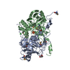
Mass: 65.409 Da / Num. of mol.: 2 / Source method: obtained synthetically / Formula: Zn
#3: Chemical
ChemComp-CL / CHLORIDE ION


Mass: 35.453 Da / Num. of mol.: 4 / Source method: obtained synthetically / Formula: Cl
#4: Chemical ChemComp-GOL / GLYCEROL / GLYCERIN / PROPANE-1,2,3-TRIOL


Mass: 92.094 Da / Num. of mol.: 1 / Source method: obtained synthetically / Formula: C3H8O3
#5: Chemical ChemComp-SO4 / SULFATE ION


Mass: 96.063 Da / Num. of mol.: 1 / Source method: obtained synthetically / Formula: SO4
#6: Water ChemComp-HOH / water


Mass: 18.015 Da / Num. of mol.: 739 / Source method: isolated from a natural source / Formula: H2O

-
Details

Sequence detailsTHE CONSTRUCT WAS EXPRESSED WITH A PURIFICATION TAG MGSDKIHHHHHHENLYFQG. THE TAG WAS REMOVED WITH ...THE CONSTRUCT WAS EXPRESSED WITH A PURIFICATION TAG MGSDKIHHHHHHENLYFQG. THE TAG WAS REMOVED WITH TEV PROTEASE LEAVING ONLY A GLYCINE (0) FOLLOWED BY THE TARGET SEQUENCE.

-
Experimental details

-
Experiment

ExperimentMethod: X-RAY DIFFRACTION / Number of used crystals: 1

-
Sample preparation

CrystalDensity Matthews: 2.16 Å3/Da / Density % sol: 43.14 %
Crystal growTemperature: 277 K / Method: vapor diffusion, sitting drop / pH: 6.9
Details: 1.7M ammonium sulfate, 2.0% polyethylene glycol 400, 15.0% Glycerol, 0.1M HEPES pH 6.9, NANODROP', VAPOR DIFFUSION, SITTING DROP, temperature 277K

-
Data collection

DiffractionMean temperature: 100 K
Diffraction sourceSource: SYNCHROTRON / Site: SSRL / Beamline: BL11-1 / Wavelength: 0.91837,0.97941,0.97904
DetectorType: MARMOSAIC 325 mm CCD / Detector: CCD / Date: May 13, 2008 / Details: Flat mirror (vertical focusing)
RadiationMonochromator: Single crystal Si(111) bent monochromator (horizontal focusing)
Protocol: MAD / Monochromatic (M) / Laue (L): M / Scattering type: x-ray
Radiation wavelength
IDWavelength (Å)Relative weight
10.918371
20.979411
30.979041
ReflectionResolution: 1.6→28.41 Å / Num. obs: 69473 / % possible obs: 98.3 % / Redundancy: 2.6 % / Biso Wilson estimate: 15.579 Å2 / Rmerge(I) obs: 0.072 / Rsym value: 0.072 / Net I/σ(I): 7
Reflection shell

Diffraction-ID: 1

Resolution (Å)Redundancy (%)Rmerge(I) obsMean I/σ(I) obsNum. measured allNum. unique allRsym value% possible all
1.6-1.642.50.4441.71217048680.44494
1.64-1.692.60.3511.61273949560.35197.7
1.69-1.742.60.3071.71247348480.30797.7
1.74-1.792.60.2433.11204346850.24397.9
1.79-1.852.60.2063.61178045750.20698.1
1.85-1.912.60.164.61139344200.1698.2
1.91-1.982.60.12951102342820.12998.3
1.98-2.072.60.1133.81061241180.11398.5
2.07-2.162.60.0997.11018139560.09998.8
2.16-2.262.60.0848.3981438120.08498.8
2.26-2.392.60.0759927535960.07599
2.39-2.532.60.0699.9885334370.06999.1
2.53-2.72.60.06510.2830432280.06599.3
2.7-2.922.60.05910.7775430100.05999.3
2.92-3.22.60.05311.6715627940.05399.6
3.2-3.582.60.04613.2638424960.04699.6
3.58-4.132.50.04712.3565122210.04799.7
4.13-5.062.50.04113.8479618980.04199.6
5.06-7.162.50.04413.8372914690.04499.8
7.16-28.412.50.04611.919838040.04697.9

-
Phasing

PhasingMethod: MAD

-
Processing

Software
NameVersionClassificationNB
REFMAC5.2.0019refinement
PHENIXrefinement
SHELXphasing
MolProbity3beta29model building
SCALAdata scaling
PDB_EXTRACT3.004data extraction
MOSFLMdata reduction
SHELXDphasing
autoSHARPphasing
RefinementMethod to determine structure: MAD / Resolution: 1.6→28.41 Å / Cor.coef. Fo:Fc: 0.973 / Cor.coef. Fo:Fc free: 0.959 / SU B: 1.367 / SU ML: 0.049 / Cross valid method: THROUGHOUT / σ(F): 0 / ESU R: 0.077 / ESU R Free: 0.079
Stereochemistry target values: MAXIMUM LIKELIHOOD WITH PHASES
Details: 1. HYDROGENS HAVE BEEN ADDED IN THE RIDING POSITIONS. 2. A MET-INHIBITION PROTOCOL WAS USED FOR SELENOMETHIONINE INCORPORATION DURING PROTEIN EXPRESSION. THE OCCUPANCY OF THE SE ATOMS IN THE ...Details: 1. HYDROGENS HAVE BEEN ADDED IN THE RIDING POSITIONS. 2. A MET-INHIBITION PROTOCOL WAS USED FOR SELENOMETHIONINE INCORPORATION DURING PROTEIN EXPRESSION. THE OCCUPANCY OF THE SE ATOMS IN THE MSE RESIDUES WAS REDUCED TO 0.75 FOR THE REDUCED SCATTERING POWER DUE TO PARTIAL S-MET INCORPORATION. 3. ZN IS MODELED BASED ON AN X-RAY FLOURESCENCE SCAN, ANOMALOUS DIFFERENCE FOURIERS, AND COORDINATION GEOMETRY. 4. A SULFATE (SO4) ION, A GLYCEROL (GOL) MOLECULE AND FOUR CHLORIDE IONS ARE MODELED BASED ON CRYSTALLIZATION CONDITIONS, ELECTRON DENSITY AND COORDINATION GEOMETRY.
RfactorNum. reflection% reflectionSelection details
Rfree0.175 3499 5 %RANDOM
Rwork0.141 ---
obs0.142 69445 98.15 %-
Solvent computationIon probe radii: 0.8 Å / Shrinkage radii: 0.8 Å / VDW probe radii: 1.2 Å / Solvent model: MASK
Displacement parametersBiso mean: 12.623 Å2
Baniso -1Baniso -2Baniso -3
1-0.03 Å20 Å20.24 Å2
2--0.1 Å20 Å2
3---0.02 Å2
Refinement stepCycle: LAST / Resolution: 1.6→28.41 Å
ProteinNucleic acidLigandSolventTotal
Num. atoms4245 0 17 739 5001
Refine LS restraints
Refine-IDTypeDev idealDev ideal targetNumber
X-RAY DIFFRACTIONr_bond_refined_d0.0140.0214487
X-RAY DIFFRACTIONr_bond_other_d0.0020.023033
X-RAY DIFFRACTIONr_angle_refined_deg1.5391.9586134
X-RAY DIFFRACTIONr_angle_other_deg0.98937358
X-RAY DIFFRACTIONr_dihedral_angle_1_deg6.1275586
X-RAY DIFFRACTIONr_dihedral_angle_2_deg32.06622.762210
X-RAY DIFFRACTIONr_dihedral_angle_3_deg11.90415672
X-RAY DIFFRACTIONr_dihedral_angle_4_deg15.4171538
X-RAY DIFFRACTIONr_chiral_restr0.0950.2659
X-RAY DIFFRACTIONr_gen_planes_refined0.0070.025142
X-RAY DIFFRACTIONr_gen_planes_other0.0010.02958
X-RAY DIFFRACTIONr_nbd_refined0.2050.2925
X-RAY DIFFRACTIONr_nbd_other0.210.23382
X-RAY DIFFRACTIONr_nbtor_refined0.1780.22163
X-RAY DIFFRACTIONr_nbtor_other0.0880.22323
X-RAY DIFFRACTIONr_xyhbond_nbd_refined0.1730.2562
X-RAY DIFFRACTIONr_xyhbond_nbd_other0.1450.21
X-RAY DIFFRACTIONr_symmetry_vdw_refined0.2170.215
X-RAY DIFFRACTIONr_symmetry_vdw_other0.3060.230
X-RAY DIFFRACTIONr_symmetry_hbond_refined0.1950.227
X-RAY DIFFRACTIONr_mcbond_it1.61632862
X-RAY DIFFRACTIONr_mcbond_other0.5731141
X-RAY DIFFRACTIONr_mcangle_it2.30254521
X-RAY DIFFRACTIONr_scbond_it3.31781809
X-RAY DIFFRACTIONr_scangle_it4.587111595
Refine LS restraints NCS

Dom-ID: 1 / Auth asym-ID: A / Ens-ID: 1 / Number: 3571 / Refine-ID: X-RAY DIFFRACTION

TypeRms dev position (Å)Weight position
MEDIUM POSITIONAL0.180.5
MEDIUM THERMAL0.812
LS refinement shellResolution: 1.6→1.642 Å / Total num. of bins used: 20
RfactorNum. reflection% reflection
Rfree0.258 244 -
Rwork0.209 4622 -
all-4866 -
obs--93.83 %

+
About Yorodumi

-
News

-
Feb 9, 2022. New format data for meta-information of EMDB entries

New format data for meta-information of EMDB entries

  • Version 3 of the EMDB header file is now the official format.
  • The previous official version 1.9 will be removed from the archive.

Related info.:EMDB header

External links:wwPDB to switch to version 3 of the EMDB data model

-
Aug 12, 2020. Covid-19 info

Covid-19 info

URL: https://pdbjlvh1.pdbj.org/emnavi/covid19.php

New page: Covid-19 featured information page in EM Navigator.

Related info.:Covid-19 info / Mar 5, 2020. Novel coronavirus structure data

+
Mar 5, 2020. Novel coronavirus structure data

Novel coronavirus structure data

Related info.:Yorodumi Speices / Aug 12, 2020. Covid-19 info

External links:COVID-19 featured content - PDBj / Molecule of the Month (242):Coronavirus Proteases

+
Jan 31, 2019. EMDB accession codes are about to change! (news from PDBe EMDB page)

EMDB accession codes are about to change! (news from PDBe EMDB page)

  • The allocation of 4 digits for EMDB accession codes will soon come to an end. Whilst these codes will remain in use, new EMDB accession codes will include an additional digit and will expand incrementally as the available range of codes is exhausted. The current 4-digit format prefixed with “EMD-” (i.e. EMD-XXXX) will advance to a 5-digit format (i.e. EMD-XXXXX), and so on. It is currently estimated that the 4-digit codes will be depleted around Spring 2019, at which point the 5-digit format will come into force.
  • The EM Navigator/Yorodumi systems omit the EMD- prefix.

Related info.:Q: What is EMD? / ID/Accession-code notation in Yorodumi/EM Navigator

External links:EMDB Accession Codes are Changing Soon! / Contact to PDBj

+
Jul 12, 2017. Major update of PDB

Major update of PDB

  • wwPDB released updated PDB data conforming to the new PDBx/mmCIF dictionary.
  • This is a major update changing the version number from 4 to 5, and with Remediation, in which all the entries are updated.
  • In this update, many items about electron microscopy experimental information are reorganized (e.g. em_software).
  • Now, EM Navigator and Yorodumi are based on the updated data.

External links:wwPDB Remediation / Enriched Model Files Conforming to OneDep Data Standards Now Available in the PDB FTP Archive

-
Yorodumi

Thousand views of thousand structures

  • Yorodumi is a browser for structure data from EMDB, PDB, SASBDB, etc.
  • This page is also the successor to EM Navigator detail page, and also detail information page/front-end page for Omokage search.
  • The word "yorodu" (or yorozu) is an old Japanese word meaning "ten thousand". "mi" (miru) is to see.

Related info.:EMDB / PDB / SASBDB / Comparison of 3 databanks / Yorodumi Search / Aug 31, 2016. New EM Navigator & Yorodumi / Yorodumi Papers / Jmol/JSmol / Function and homology information / Changes in new EM Navigator and Yorodumi

Read more