 Movie
Movie Controller

[English] 日本語
 Yorodumi
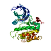
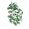
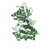
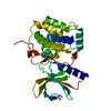
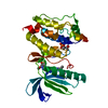
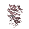
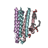
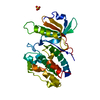
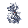
- PDB-3dbd: Crystal structure of an activated (Thr->Asp) Polo-like kinase 1 (... -
+ Open data

- Basic information
| Entry | Database: PDB / ID: 3dbd | ||||||
|---|---|---|---|---|---|---|---|
| Title | Crystal structure of an activated (Thr->Asp) Polo-like kinase 1 (Plk1) catalytic domain in complex with Compound 094 | ||||||
 Components Components | Polo-like kinase 1 | ||||||
 Keywords Keywords | TRANSFERASE / Polo-like kinase 1 / Plk1 / catalytic domain / small-molecule inhibitor / Kinase | ||||||
| Function / homology |  Function and homology information Function and homology informationPhosphorylation of Emi1 / Condensation of Prophase Chromosomes / Resolution of Sister Chromatid Cohesion / Regulation of PLK1 Activity at G2/M Transition / Mitotic Metaphase/Anaphase Transition / Mitotic Telophase/Cytokinesis / EML4 and NUDC in mitotic spindle formation / Polo-like kinase mediated events / Cyclin A/B1/B2 associated events during G2/M transition / The role of GTSE1 in G2/M progression after G2 checkpoint ...Phosphorylation of Emi1 / Condensation of Prophase Chromosomes / Resolution of Sister Chromatid Cohesion / Regulation of PLK1 Activity at G2/M Transition / Mitotic Metaphase/Anaphase Transition / Mitotic Telophase/Cytokinesis / EML4 and NUDC in mitotic spindle formation / Polo-like kinase mediated events / Cyclin A/B1/B2 associated events during G2/M transition / The role of GTSE1 in G2/M progression after G2 checkpoint / polo kinase / mitotic spindle organization / kinetochore / spindle pole / mitotic cell cycle / retina development in camera-type eye / midbody / cell division / protein serine/threonine kinase activity / centrosome / ATP binding / nucleus / cytoplasm Similarity search - Function | ||||||
| Biological species |  | ||||||
| Method |  X-RAY DIFFRACTION / SYNCHROTRON / MOLECULAR REPLACEMENT / Resolution: 3.05 Å X-RAY DIFFRACTION / SYNCHROTRON / MOLECULAR REPLACEMENT / Resolution: 3.05 Å | ||||||
 Authors Authors | Elling, R.A. / Barr, K.J. / Romanowski, M.J. | ||||||
 Citation Citation | Journal: Bioorg.Med.Chem.Lett. / Year: 2008 Title: Design and synthesis of 2-amino-pyrazolopyridines as Polo-like kinase 1 inhibitors. Authors: Fucini, R.V. / Hanan, E.J. / Romanowski, M.J. / Elling, R.A. / Lew, W. / Barr, K.J. / Zhu, J. / Yoburn, J.C. / Liu, Y. / Fahr, B.T. / Fan, J. / Lu, Y. / Pham, P. / Choong, I.C. / ...Authors: Fucini, R.V. / Hanan, E.J. / Romanowski, M.J. / Elling, R.A. / Lew, W. / Barr, K.J. / Zhu, J. / Yoburn, J.C. / Liu, Y. / Fahr, B.T. / Fan, J. / Lu, Y. / Pham, P. / Choong, I.C. / VanderPorten, E.C. / Bui, M. / Purkey, H.E. / Evanchik, M.J. / Yang, W. | ||||||
| History |
|
- Structure visualization
| Structure viewer | Molecule: MolmilJmol/JSmol |
|---|
- Downloads & links
-Download
| PDBx/mmCIF format | 3dbd.cif.gz | 66.6 KB | Display | PDBx/mmCIF format |
|---|---|---|---|---|
| PDB format | pdb3dbd.ent.gz | 48.7 KB | Display | PDB format |
| PDBx/mmJSON format | 3dbd.json.gz | Tree view | PDBx/mmJSON format | |
| Others |  Other downloads Other downloads |
-Validation report
| Arichive directory | https://data.pdbj.org/pub/pdb/validation_reports/db/3dbdftp://data.pdbj.org/pub/pdb/validation_reports/db/3dbd | HTTPS FTP |
|---|
-Related structure data
| Related structure data | 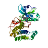 3dbcC  3dbeC 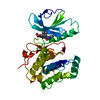 3dbfC  3d5wS S: Starting model for refinement C: citing same article ( |
|---|---|
| Similar structure data |


-Links
 PDBj
PDBj- Assembly
| Deposited unit | 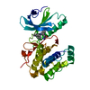
| ||||||||
|---|---|---|---|---|---|---|---|---|---|
| 1 |
| ||||||||
| Unit cell |
|
-Components
| #1: Protein | Mass: 35992.953 Da / Num. of mol.: 1 / Fragment: Plk1 kinase domain / Mutation: T196D Source method: isolated from a genetically manipulated source Source: (gene. exp.)  |
|---|---|
| #2: Chemical | ChemComp-3FR / 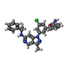  Mass: 481.976 Da / Num. of mol.: 1 / Source method: obtained synthetically / Formula: C28H24ClN5O Mass: 481.976 Da / Num. of mol.: 1 / Source method: obtained synthetically / Formula: C28H24ClN5O |
| #3: Water | ChemComp-HOH /  Mass: 18.015 Da / Num. of mol.: 2 / Source method: isolated from a natural source / Formula: H2O Mass: 18.015 Da / Num. of mol.: 2 / Source method: isolated from a natural source / Formula: H2O |
-Experimental details
-Experiment
| Experiment | Method: X-RAY DIFFRACTION / Number of used crystals: 1 |
|---|
- Sample preparation
| Crystal | Density Matthews: 2.87 Å3/Da / Density % sol: 57.07 % |
|---|---|
| Crystal grow | Temperature: 277 K / Method: vapor diffusion, hanging drop / pH: 5.6 Details: hanging-drop vapor diffusion at 4 C (277K); protein at 7 mg/ml in 50 mM Tris-HCl pH 7.5, 200 mM NaCl and 3 mM DTT; crystallization condition: 0.1 M tri-sodium citrate pH 5.6, 0.2 M (NH4) ...Details: hanging-drop vapor diffusion at 4 C (277K); protein at 7 mg/ml in 50 mM Tris-HCl pH 7.5, 200 mM NaCl and 3 mM DTT; crystallization condition: 0.1 M tri-sodium citrate pH 5.6, 0.2 M (NH4)2SO4, and 25% PEG 3350; cryoprotectant: 15% ethylene glycol., VAPOR DIFFUSION, HANGING DROP |
-Data collection
| Diffraction | Mean temperature: 160 K |
|---|---|
| Diffraction source | Source: SYNCHROTRON / Site: SSRL  / Beamline: BL9-1 / Wavelength: 0.98 Å / Beamline: BL9-1 / Wavelength: 0.98 Å |
| Detector | Type: ADSC QUANTUM 315 / Detector: CCD / Date: Feb 8, 2007 |
| Radiation | Monochromator: Side-scattering cube root I-beam bent single crystal; asymmetric cut 12.2 degs. Crystal type: Si(311) Protocol: SINGLE WAVELENGTH / Monochromatic (M) / Laue (L): M / Scattering type: x-ray |
| Radiation wavelength | Wavelength: 0.98 Å / Relative weight: 1 |
| Reflection | Resolution: 3.05→30 Å / Num. obs: 7979 / % possible obs: 99.7 % / Redundancy: 3.5 % / Rmerge(I) obs: 0.073 / Net I/σ(I): 8.5 |
| Reflection shell | Resolution: 3.05→3.21 Å / Rmerge(I) obs: 0.364 / Mean I/σ(I) obs: 2.1 / % possible all: 100 |
- Processing
| Software |
| ||||||||||||||||||||||||||||||||||||||||||||||||||||||||||||||||||||||||||||||||||||||||||||||||||||||||||||||||||||||||||||||||||||||||||||||||||||||||||||||||||||||||||
|---|---|---|---|---|---|---|---|---|---|---|---|---|---|---|---|---|---|---|---|---|---|---|---|---|---|---|---|---|---|---|---|---|---|---|---|---|---|---|---|---|---|---|---|---|---|---|---|---|---|---|---|---|---|---|---|---|---|---|---|---|---|---|---|---|---|---|---|---|---|---|---|---|---|---|---|---|---|---|---|---|---|---|---|---|---|---|---|---|---|---|---|---|---|---|---|---|---|---|---|---|---|---|---|---|---|---|---|---|---|---|---|---|---|---|---|---|---|---|---|---|---|---|---|---|---|---|---|---|---|---|---|---|---|---|---|---|---|---|---|---|---|---|---|---|---|---|---|---|---|---|---|---|---|---|---|---|---|---|---|---|---|---|---|---|---|---|---|---|---|---|---|
| Refinement | Method to determine structure: MOLECULAR REPLACEMENT Starting model: 3d5w Resolution: 3.05→30 Å / Cor.coef. Fo:Fc: 0.896 / Cor.coef. Fo:Fc free: 0.871 / SU B: 22.309 / SU ML: 0.385 / Cross valid method: THROUGHOUT / ESU R Free: 0.472 / Stereochemistry target values: MAXIMUM LIKELIHOOD
| ||||||||||||||||||||||||||||||||||||||||||||||||||||||||||||||||||||||||||||||||||||||||||||||||||||||||||||||||||||||||||||||||||||||||||||||||||||||||||||||||||||||||||
| Solvent computation | Ion probe radii: 0.8 Å / Shrinkage radii: 0.8 Å / VDW probe radii: 1.2 Å / Solvent model: BABINET MODEL WITH MASK | ||||||||||||||||||||||||||||||||||||||||||||||||||||||||||||||||||||||||||||||||||||||||||||||||||||||||||||||||||||||||||||||||||||||||||||||||||||||||||||||||||||||||||
| Displacement parameters | Biso mean: 58.728 Å2 | ||||||||||||||||||||||||||||||||||||||||||||||||||||||||||||||||||||||||||||||||||||||||||||||||||||||||||||||||||||||||||||||||||||||||||||||||||||||||||||||||||||||||||
| Refinement step | Cycle: LAST / Resolution: 3.05→30 Å
| ||||||||||||||||||||||||||||||||||||||||||||||||||||||||||||||||||||||||||||||||||||||||||||||||||||||||||||||||||||||||||||||||||||||||||||||||||||||||||||||||||||||||||
| Refine LS restraints |
| ||||||||||||||||||||||||||||||||||||||||||||||||||||||||||||||||||||||||||||||||||||||||||||||||||||||||||||||||||||||||||||||||||||||||||||||||||||||||||||||||||||||||||
| LS refinement shell | Resolution: 3.05→3.157 Å / Total num. of bins used: 15
|
