 Movie
Movie Controller

[English] 日本語
 Yorodumi
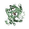
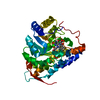
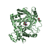
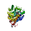
- PDB-3d4u: Bovine thrombin-activatable fibrinolysis inhibitor (TAFIa) in com... -
+ Open data

- Basic information
| Entry | Database: PDB / ID: 3d4u | ||||||
|---|---|---|---|---|---|---|---|
| Title | Bovine thrombin-activatable fibrinolysis inhibitor (TAFIa) in complex with tick-derived carboxypeptidase inhibitor. | ||||||
 Components Components | (Carboxypeptidase ...) x 2 | ||||||
 Keywords Keywords | HYDROLASE/HYDROLASE INHIBITOR / protease-inhibitor complex / Carboxypeptidase / Glycoprotein / Hydrolase / Metal-binding / Metalloprotease / Protease / Secreted / Zymogen / Blood coagulation / Fibrinolysis / Metalloenzyme inhibitor / Metalloprotease inhibitor / HYDROLASE-HYDROLASE INHIBITOR COMPLEX | ||||||
| Function / homology |  Function and homology information Function and homology informationRegulation of Complement cascade / Metabolism of Angiotensinogen to Angiotensins / carboxypeptidase U / acquisition of nutrients from host / metalloendopeptidase inhibitor activity / fibrinolysis / metallocarboxypeptidase activity / enzyme inhibitor activity / blood coagulation / toxin activity ...Regulation of Complement cascade / Metabolism of Angiotensinogen to Angiotensins / carboxypeptidase U / acquisition of nutrients from host / metalloendopeptidase inhibitor activity / fibrinolysis / metallocarboxypeptidase activity / enzyme inhibitor activity / blood coagulation / toxin activity / proteolysis / : / extracellular region / zinc ion binding Similarity search - Function | ||||||
| Biological species |  Rhipicephalus bursa (arthropod) Rhipicephalus bursa (arthropod) | ||||||
| Method |  X-RAY DIFFRACTION / SYNCHROTRON / MOLECULAR REPLACEMENT / Resolution: 1.7 Å X-RAY DIFFRACTION / SYNCHROTRON / MOLECULAR REPLACEMENT / Resolution: 1.7 Å | ||||||
 Authors Authors | Sanglas, L. / Valnickova, Z. / Arolas, J.L. / Pallares, I. / Guevara, T. / Sola, M. / Kristensen, T. / Enghild, J.J. / Aviles, F.X. / Gomis-Ruth, F.X. | ||||||
 Citation Citation | Journal: Mol.Cell / Year: 2008 Title: Structure of activated thrombin-activatable fibrinolysis inhibitor, a molecular link between coagulation and fibrinolysis. Authors: Sanglas, L. / Valnickova, Z. / Arolas, J.L. / Pallares, I. / Guevara, T. / Sola, M. / Kristensen, T. / Enghild, J.J. / Aviles, F.X. / Gomis-Ruth, F.X. #1: Journal: J.Mol.Biol. / Year: 2002Title: Human procarboxypeptidase B: three-dimensional structure and implications for thrombin-activatable fibrinolysis inhibitor (TAFI) Authors: Barbosa Pereira, P.J. / Oliva, B. / Ferrer-Orta, C. / Coll, M. / Vendrell, J. | ||||||
| History |
|
- Structure visualization
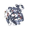
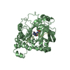
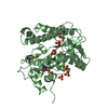
| Structure viewer | Molecule: MolmilJmol/JSmol |
|---|
- Downloads & links
-Download
| PDBx/mmCIF format | 3d4u.cif.gz | 99.1 KB | Display | PDBx/mmCIF format |
|---|---|---|---|---|
| PDB format | pdb3d4u.ent.gz | 74.3 KB | Display | PDB format |
| PDBx/mmJSON format | 3d4u.json.gz | Tree view | PDBx/mmJSON format | |
| Others |  Other downloads Other downloads |
-Validation report
| Arichive directory | https://data.pdbj.org/pub/pdb/validation_reports/d4/3d4uftp://data.pdbj.org/pub/pdb/validation_reports/d4/3d4u | HTTPS FTP |
|---|
-Related structure data
| Related structure data | 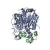 1zliS S: Starting model for refinement |
|---|---|
| Similar structure data |


-Links
 PDBj
PDBj
- Assembly
| Deposited unit | 
| ||||||||
|---|---|---|---|---|---|---|---|---|---|
| 1 |
| ||||||||
| Unit cell |
|
-Components
-Carboxypeptidase ... , 2 types, 2 molecules AB
| #1: Protein | Mass: 36029.906 Da / Num. of mol.: 1 / Source method: isolated from a natural source / Source: (natural) |
|---|---|
| #2: Protein | Mass: 7806.957 Da / Num. of mol.: 1 Source method: isolated from a genetically manipulated source Source: (gene. exp.) Rhipicephalus bursa (arthropod) / Plasmid: pBAT-4-OmpA-TCI / Production host:  |
-Non-polymers , 4 types, 366 molecules 


| #3: Chemical | ChemComp-ZN /  Mass: 65.409 Da / Num. of mol.: 1 / Source method: obtained synthetically / Formula: Zn Mass: 65.409 Da / Num. of mol.: 1 / Source method: obtained synthetically / Formula: Zn | ||||
|---|---|---|---|---|---|
| #4: Chemical |  Mass: 59.044 Da / Num. of mol.: 2 / Source method: obtained synthetically / Formula: C2H3O2 Mass: 59.044 Da / Num. of mol.: 2 / Source method: obtained synthetically / Formula: C2H3O2#5: Chemical | ChemComp-SO4 /  Mass: 96.063 Da / Num. of mol.: 5 / Source method: obtained synthetically / Formula: SO4 Mass: 96.063 Da / Num. of mol.: 5 / Source method: obtained synthetically / Formula: SO4#6: Water | ChemComp-HOH / | Mass: 18.015 Da / Num. of mol.: 358 / Source method: isolated from a natural source / Formula: H2O |
-Details
| Has protein modification | Y |
|---|
-Experimental details
-Experiment
| Experiment | Method: X-RAY DIFFRACTION / Number of used crystals: 1 |
|---|
- Sample preparation
| Crystal | Density Matthews: 2.98 Å3/Da / Density % sol: 58.67 % |
|---|---|
| Crystal grow | Temperature: 293 K / Method: vapor diffusion, sitting drop / pH: 4.6 Details: 0.2M (NH4)2SO4, 0.1M NaAcO, 10% PEG 4000, pH 4.6, VAPOR DIFFUSION, SITTING DROP, temperature 293K |
-Data collection
| Diffraction | Mean temperature: 100 K |
|---|---|
| Diffraction source | Source: SYNCHROTRON / Site: ESRF  / Beamline: ID23-2 / Wavelength: 0.8726 Å / Beamline: ID23-2 / Wavelength: 0.8726 Å |
| Detector | Type: MAR CCD 165 mm / Detector: CCD / Date: Jul 18, 2007 Details: horizontally diffracting Si (111) monochromator and Pt coated mirrors in a Kirkpatrick-Baez (KB) geometry as the focusing system. |
| Radiation | Monochromator: Horizontally diffracting Si (111) monochromator and Pt coated mirrors in a Kirkpatrick-Baez (KB) geometry as the focusing system Protocol: SINGLE WAVELENGTH / Monochromatic (M) / Laue (L): M / Scattering type: x-ray |
| Radiation wavelength | Wavelength: 0.8726 Å / Relative weight: 1 |
| Reflection | Resolution: 1.7→48.1 Å / Num. all: 62124 / Num. obs: 57962 / % possible obs: 93.3 % / Observed criterion σ(F): 0 / Observed criterion σ(I): 0 / Redundancy: 15.1 % / Biso Wilson estimate: 18.6 Å2 / Rmerge(I) obs: 0.099 / Rsym value: 0.099 / Net I/σ(I): 20.6 |
| Reflection shell | Resolution: 1.7→1.79 Å / Redundancy: 9.2 % / Rmerge(I) obs: 0.611 / Mean I/σ(I) obs: 9.2 / Num. unique all: 8032 / Rsym value: 0.611 / % possible all: 95.5 |
- Processing
| Software |
| ||||||||||||||||||||||||||||||||||||||||||||||||||||||||||||||||||||||||||||||||||||||||||||||||||||
|---|---|---|---|---|---|---|---|---|---|---|---|---|---|---|---|---|---|---|---|---|---|---|---|---|---|---|---|---|---|---|---|---|---|---|---|---|---|---|---|---|---|---|---|---|---|---|---|---|---|---|---|---|---|---|---|---|---|---|---|---|---|---|---|---|---|---|---|---|---|---|---|---|---|---|---|---|---|---|---|---|---|---|---|---|---|---|---|---|---|---|---|---|---|---|---|---|---|---|---|---|---|
| Refinement | Method to determine structure: MOLECULAR REPLACEMENT Starting model: 1ZLI Resolution: 1.7→48.08 Å / Cor.coef. Fo:Fc: 0.966 / Cor.coef. Fo:Fc free: 0.968 / SU B: 3.053 / SU ML: 0.046 / TLS residual ADP flag: LIKELY RESIDUAL / Cross valid method: THROUGHOUT / σ(F): 0 / σ(I): 0 / ESU R: 0.074 / ESU R Free: 0.071 / Stereochemistry target values: MAXIMUM LIKELIHOOD / Details: HYDROGENS HAVE BEEN ADDED IN THE RIDING POSITIONS
| ||||||||||||||||||||||||||||||||||||||||||||||||||||||||||||||||||||||||||||||||||||||||||||||||||||
| Solvent computation | Ion probe radii: 0.8 Å / Shrinkage radii: 0.8 Å / VDW probe radii: 1.4 Å / Solvent model: BABINET MODEL WITH MASK | ||||||||||||||||||||||||||||||||||||||||||||||||||||||||||||||||||||||||||||||||||||||||||||||||||||
| Displacement parameters | Biso mean: 20.858 Å2
| ||||||||||||||||||||||||||||||||||||||||||||||||||||||||||||||||||||||||||||||||||||||||||||||||||||
| Refinement step | Cycle: LAST / Resolution: 1.7→48.08 Å
| ||||||||||||||||||||||||||||||||||||||||||||||||||||||||||||||||||||||||||||||||||||||||||||||||||||
| Refine LS restraints |
| ||||||||||||||||||||||||||||||||||||||||||||||||||||||||||||||||||||||||||||||||||||||||||||||||||||
| LS refinement shell | Resolution: 1.7→1.744 Å / Total num. of bins used: 20
| ||||||||||||||||||||||||||||||||||||||||||||||||||||||||||||||||||||||||||||||||||||||||||||||||||||
| Refinement TLS params. | Method: refined / Refine-ID: X-RAY DIFFRACTION
| ||||||||||||||||||||||||||||||||||||||||||||||||||||||||||||||||||||||||||||||||||||||||||||||||||||
| Refinement TLS group |
|
