 Movie
Movie Controller

[English] 日本語
 Yorodumi
- PDB-1ujm: Crystal structure of aldehyde reductase 2 from Sporobolomyces sal... -
+ Open data

- Basic information
| Entry | Database: PDB / ID: 1ujm | ||||||
|---|---|---|---|---|---|---|---|
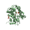
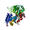
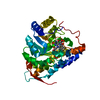
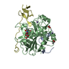
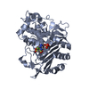
| Title | Crystal structure of aldehyde reductase 2 from Sporobolomyces salmonicolor AKU4429 | ||||||
 Components Components | Aldehyde reductase II | ||||||
 Keywords Keywords | OXIDOREDUCTASE / NADPH-dependent enzyme | ||||||
| Function / homology |  Function and homology information Function and homology information | ||||||
| Biological species |  Sporidiobolus salmonicolor (yeast) Sporidiobolus salmonicolor (yeast) | ||||||
| Method |  X-RAY DIFFRACTION / SYNCHROTRON / MAD / Resolution: 2 Å X-RAY DIFFRACTION / SYNCHROTRON / MAD / Resolution: 2 Å | ||||||
 Authors Authors | Kamitori, S. / Iguchi, A. / Ohtaki, A. / Kita, K. | ||||||
 Citation Citation | Journal: To be Published Title: Crystal structure of aldehyde reductase 2 from Sporobolomyces salmonicolor AKU4429 at 2.0 A resolution Authors: Kamitori, S. / Iguchi, A. / Ohtaki, A. / Kita, K. | ||||||
| History |
|
- Structure visualization
| Structure viewer | Molecule: MolmilJmol/JSmol |
|---|
- Downloads & links
-Download
| PDBx/mmCIF format | 1ujm.cif.gz | 158.2 KB | Display | PDBx/mmCIF format |
|---|---|---|---|---|
| PDB format | pdb1ujm.ent.gz | 122.9 KB | Display | PDB format |
| PDBx/mmJSON format | 1ujm.json.gz | Tree view | PDBx/mmJSON format | |
| Others |  Other downloads Other downloads |
-Validation report
| Arichive directory | https://data.pdbj.org/pub/pdb/validation_reports/uj/1ujmftp://data.pdbj.org/pub/pdb/validation_reports/uj/1ujm | HTTPS FTP |
|---|
-Related structure data
| Similar structure data |
|---|


-Links
 PDBj
PDBj- Assembly
| Deposited unit | 
| ||||||||
|---|---|---|---|---|---|---|---|---|---|
| 1 | 
| ||||||||
| 2 | 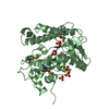
| ||||||||
| Unit cell |
|
-Components
| #1: Protein | Mass: 37459.668 Da / Num. of mol.: 2 Source method: isolated from a genetically manipulated source Source: (gene. exp.) Sporidiobolus salmonicolor (yeast) / Strain: AKU4429 / Plasmid: pUC / Production host:  #2: Chemical | ChemComp-SO4 /   Mass: 96.063 Da / Num. of mol.: 10 / Source method: obtained synthetically / Formula: SO4 Mass: 96.063 Da / Num. of mol.: 10 / Source method: obtained synthetically / Formula: SO4#3: Water | ChemComp-HOH / |  Mass: 18.015 Da / Num. of mol.: 649 / Source method: isolated from a natural source / Formula: H2O Mass: 18.015 Da / Num. of mol.: 649 / Source method: isolated from a natural source / Formula: H2OHas protein modification | Y | |
|---|
-Experimental details
-Experiment
| Experiment | Method: X-RAY DIFFRACTION / Number of used crystals: 1 |
|---|
- Sample preparation
| Crystal | Density Matthews: 2.04 Å3/Da / Density % sol: 39.29 % |
|---|---|
| Crystal grow | Temperature: 277 K / Method: vapor diffusion, hanging drop / pH: 4.6 Details: 30% PEG MME 2000, 0.1M Na Acetate, 0.2M Ammonium sulfate, pH 4.6, VAPOR DIFFUSION, HANGING DROP, temperature 277K |
-Data collection
| Diffraction | Mean temperature: 173 K | ||||||||||||
|---|---|---|---|---|---|---|---|---|---|---|---|---|---|
| Diffraction source | Source: SYNCHROTRON / Site: Photon Factory  / Beamline: AR-NW12A / Wavelength: 0.97899, 0.97916, 1.0 / Beamline: AR-NW12A / Wavelength: 0.97899, 0.97916, 1.0 | ||||||||||||
| Detector | Type: ADSC QUANTUM 4 / Detector: CCD / Date: Jun 28, 2003 | ||||||||||||
| Radiation | Protocol: MAD / Monochromatic (M) / Laue (L): M / Scattering type: x-ray | ||||||||||||
| Radiation wavelength |
| ||||||||||||
| Reflection | Resolution: 2→39.03 Å / Num. all: 47081 / Num. obs: 47081 / % possible obs: 99.9 % / Observed criterion σ(F): 0 / Observed criterion σ(I): 0 / Redundancy: 7.2 % / Biso Wilson estimate: 12.3 Å2 / Rmerge(I) obs: 0.059 / Rsym value: 0.055 / Net I/σ(I): 8 | ||||||||||||
| Reflection shell | Resolution: 2→2.11 Å / Redundancy: 7.2 % / Rmerge(I) obs: 0.104 / Mean I/σ(I) obs: 6.3 / Num. unique all: 6783 / Rsym value: 0.096 / % possible all: 99.9 |
- Processing
| Software |
| |||||||||||||||||||||||||
|---|---|---|---|---|---|---|---|---|---|---|---|---|---|---|---|---|---|---|---|---|---|---|---|---|---|---|
| Refinement | Method to determine structure: MAD / Resolution: 2→39.03 Å / Rfactor Rfree error: 0.004 / Data cutoff high absF: 2323120.14 / Data cutoff low absF: 0 / Isotropic thermal model: RESTRAINED / Cross valid method: THROUGHOUT / σ(F): 0 / σ(I): 0 / Stereochemistry target values: CNS1.1
| |||||||||||||||||||||||||
| Solvent computation | Solvent model: FLAT MODEL / Bsol: 56.1516 Å2 / ksol: 0.359713 e/Å3 | |||||||||||||||||||||||||
| Displacement parameters | Biso mean: 24.4 Å2
| |||||||||||||||||||||||||
| Refine analyze |
| |||||||||||||||||||||||||
| Refinement step | Cycle: LAST / Resolution: 2→39.03 Å
| |||||||||||||||||||||||||
| Refine LS restraints |
| |||||||||||||||||||||||||
| LS refinement shell | Resolution: 2→2.13 Å / Rfactor Rfree error: 0.01 / Total num. of bins used: 6
| |||||||||||||||||||||||||
| Xplor file |
|
