[English] 日本語
Yorodumi
- PDB-3d4s: Cholesterol bound form of human beta2 adrenergic receptor. -

+
Open data


ID or keywords:

Loading...

-
Basic information

Entry
Database: PDB / ID: 3d4s
TitleCholesterol bound form of human beta2 adrenergic receptor.
ComponentsBeta-2 adrenergic receptor/T4-lysozyme chimera
KeywordsMEMBRANE PROTEIN / GPCR / Lysozyme / Fusion / Adrenergic / timolol / G-protein coupled receptor / Glycoprotein / Lipoprotein / Palmitate / Phosphoprotein / Receptor / Transducer / Transmembrane / Structural Genomics / PSI-2 / Protein Structure Initiative / Accelerated Technologies Center for Gene to 3D Structure / ATCG3D / GPCR Network
Function / homology
Function and homology information


desensitization of G protein-coupled receptor signaling pathway by arrestin / beta2-adrenergic receptor activity / norepinephrine-epinephrine-mediated vasodilation involved in regulation of systemic arterial blood pressure / positive regulation of mini excitatory postsynaptic potential / positive regulation of cAMP-dependent protein kinase activity / norepinephrine binding / Adrenoceptors / heat generation / positive regulation of autophagosome maturation / positive regulation of AMPA receptor activity ...desensitization of G protein-coupled receptor signaling pathway by arrestin / beta2-adrenergic receptor activity / norepinephrine-epinephrine-mediated vasodilation involved in regulation of systemic arterial blood pressure / positive regulation of mini excitatory postsynaptic potential / positive regulation of cAMP-dependent protein kinase activity / norepinephrine binding / Adrenoceptors / heat generation / positive regulation of autophagosome maturation / positive regulation of AMPA receptor activity / activation of transmembrane receptor protein tyrosine kinase activity / negative regulation of smooth muscle contraction / positive regulation of lipophagy / response to psychosocial stress / negative regulation of multicellular organism growth / endosome to lysosome transport / adrenergic receptor signaling pathway / diet induced thermogenesis / neuronal dense core vesicle / positive regulation of protein kinase A signaling / adenylate cyclase binding / smooth muscle contraction / potassium channel regulator activity / positive regulation of bone mineralization / adenylate cyclase-activating adrenergic receptor signaling pathway / brown fat cell differentiation / regulation of sodium ion transport / bone resorption / viral release from host cell by cytolysis / activation of adenylate cyclase activity / receptor-mediated endocytosis / response to cold / peptidoglycan catabolic process / clathrin-coated endocytic vesicle membrane / adenylate cyclase-modulating G protein-coupled receptor signaling pathway / positive regulation of protein serine/threonine kinase activity / cellular response to amyloid-beta / cell wall macromolecule catabolic process / Cargo recognition for clathrin-mediated endocytosis / lysozyme / Clathrin-mediated endocytosis / lysozyme activity / amyloid-beta binding / positive regulation of cold-induced thermogenesis / G alpha (s) signalling events / host cell cytoplasm / positive regulation of MAPK cascade / transcription by RNA polymerase II / lysosome / cell surface receptor signaling pathway / early endosome / receptor complex / endosome membrane / Ub-specific processing proteases / endosome / defense response to bacterium / apical plasma membrane / protein-containing complex binding / Golgi apparatus / protein homodimerization activity / positive regulation of transcription by RNA polymerase II / membrane / identical protein binding / nucleus / plasma membrane
Similarity search - Function
Beta 2 adrenoceptor / Adrenoceptor family / Rhopdopsin 7-helix transmembrane proteins / Rhodopsin 7-helix transmembrane proteins / Lysozyme - #40 / Endolysin T4 type / T4-type lysozyme / Glycoside hydrolase, family 24 / Lysozyme domain superfamily / Phage lysozyme ...Beta 2 adrenoceptor / Adrenoceptor family / Rhopdopsin 7-helix transmembrane proteins / Rhodopsin 7-helix transmembrane proteins / Lysozyme - #40 / Endolysin T4 type / T4-type lysozyme / Glycoside hydrolase, family 24 / Lysozyme domain superfamily / Phage lysozyme / Serpentine type 7TM GPCR chemoreceptor Srsx / Lysozyme / G-protein coupled receptors family 1 signature. / G protein-coupled receptor, rhodopsin-like / GPCR, rhodopsin-like, 7TM / G-protein coupled receptors family 1 profile. / 7 transmembrane receptor (rhodopsin family) / Lysozyme-like domain superfamily / Up-down Bundle / Orthogonal Bundle / Mainly Alpha
Similarity search - Domain/homology
CHOLESTEROL / (2R)-2,3-dihydroxypropyl (9Z)-octadec-9-enoate / Chem-TIM / Endolysin / Beta-2 adrenergic receptor
Similarity search - Component
Biological speciesHomo sapiens (human)
Enterobacteria phage T4 (virus)
MethodX-RAY DIFFRACTION / SYNCHROTRON / MOLECULAR REPLACEMENT / Resolution: 2.8 Å
AuthorsHanson, M.A. / Cherezov, V. / Roth, C.B. / Griffith, M.T. / Jaakola, V.-P. / Chien, E.Y.T. / Velasquez, J. / Kuhn, P. / Stevens, R.C. / Accelerated Technologies Center for Gene to 3D Structure (ATCG3D) / GPCR Network (GPCR)
CitationJournal: Structure / Year: 2008
Title: A specific cholesterol binding site is established by the 2.8 A structure of the human beta2-adrenergic receptor.
Authors: Hanson, M.A. / Cherezov, V. / Griffith, M.T. / Roth, C.B. / Jaakola, V.P. / Chien, E.Y. / Velasquez, J. / Kuhn, P. / Stevens, R.C.
History
DepositionMay 14, 2008Deposition site: RCSB / Processing site: RCSB
Revision 1.0Jun 17, 2008Provider: repository / Type: Initial release
Revision 1.1Jul 13, 2011Group: Version format compliance
Revision 1.2Aug 8, 2012Group: Other
Revision 1.3Jun 24, 2015Group: Database references / Source and taxonomy
Revision 1.4Jul 5, 2017Group: Database references / Source and taxonomy / Category: entity_src_gen / struct_ref_seq / Item: _struct_ref_seq.db_align_end
Revision 1.5Jan 24, 2018Group: Structure summary / Category: audit_author / Item: _audit_author.name
Revision 1.6Oct 20, 2021Group: Database references / Derived calculations / Category: database_2 / struct_ref_seq_dif / struct_site
Item: _database_2.pdbx_DOI / _database_2.pdbx_database_accession ..._database_2.pdbx_DOI / _database_2.pdbx_database_accession / _struct_ref_seq_dif.details / _struct_site.pdbx_auth_asym_id / _struct_site.pdbx_auth_comp_id / _struct_site.pdbx_auth_seq_id
Revision 1.7Aug 30, 2023Group: Data collection / Refinement description
Category: chem_comp_atom / chem_comp_bond / pdbx_initial_refinement_model

-
Structure visualization

Structure viewerMolecule:
MolmilJmol/JSmol

Downloads & links

-
Assembly

Deposited unit
A: Beta-2 adrenergic receptor/T4-lysozyme chimera
hetero molecules


Theoretical massNumber of molelcules
Total (without water)58,1177
Polymers55,9581
Non-polymers2,1596
Water36020
1


  • Idetical with deposited unit
  • defined by author&software
TypeNameSymmetry operationNumber
identity operation1_555x,y,z1
MethodPISA
Unit cell
Length a, b, c (Å)40.000, 75.700, 172.730
Angle α, β, γ (deg.)90.00, 90.00, 90.00
Int Tables number19
Space group name H-MP212121

-
Components

#1: Protein Beta-2 adrenergic receptor/T4-lysozyme chimera / Beta-2 adrenoceptor / Beta-2 adrenoreceptor / Lysis protein / Muramidase / Endolysin


Mass: 55957.926 Da / Num. of mol.: 1 / Mutation: E122W, N187E, C1054T, C1097A
Source method: isolated from a genetically manipulated source
Source: (gene. exp.) Homo sapiens (human), (gene. exp.) Enterobacteria phage T4 (virus)
Gene: ADRB2, ADRB2R, B2AR / E / Plasmid: pFastBac / Production host: 
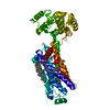
Spodoptera frugiperda (fall armyworm) / References: UniProt: P07550, UniProt: P00720
#2: Chemical ChemComp-TIM / (2S)-1-(tert-butylamino)-3-[(4-morpholin-4-yl-1,2,5-thiadiazol-3-yl)oxy]propan-2-ol / Timolol maleate / Timolol


Mass: 316.420 Da / Num. of mol.: 1 / Source method: obtained synthetically / Formula: C13H24N4O3S / Comment: medication*YM
#3: Chemical ChemComp-CLR / CHOLESTEROL / Cholesterol


Mass: 386.654 Da / Num. of mol.: 2 / Source method: obtained synthetically / Formula: C27H46O
#4: Chemical ChemComp-OLC / (2R)-2,3-dihydroxypropyl (9Z)-octadec-9-enoate / 1-Oleoyl-R-glycerol


Mass: 356.540 Da / Num. of mol.: 3 / Source method: obtained synthetically / Formula: C21H40O4
#5: Water ChemComp-HOH / water / Water


Mass: 18.015 Da / Num. of mol.: 20 / Source method: isolated from a natural source / Formula: H2O
Sequence detailsTHE STRUCTURE IS AN INTERNAL FUSION PROTEIN WITH LYSOZYME. AN OFFSET 1000 HAS BEEN ADDED TO ...THE STRUCTURE IS AN INTERNAL FUSION PROTEIN WITH LYSOZYME. AN OFFSET 1000 HAS BEEN ADDED TO ORIGINAL SEQUENCE DATABASE RESIDUE NUMBERS (2-161) OF THE LYSOZYME PART IN COORDINATES TO DISTINGUISH THE LYSOZYME PART IN THE CHAIN. THEREFORE THE RESIDUES OF LYSOZYME PART HAVE NUMBERS A1002-A1161.

-
Experimental details

-
Experiment

ExperimentMethod: X-RAY DIFFRACTION / Number of used crystals: 9

-
Sample preparation

CrystalDensity Matthews: 2.34 Å3/Da / Density % sol: 47.36 % / Description: THIS ENTRY IS A JCIMPT/ATCG3D STRUCTURE
Crystal growTemperature: 293 K / Method: mesophase / pH: 7
Details: 28% v/v PEG 400, 300mM K formate, 100mM Bis-tris propane pH 7.0, 2mM Timolol, MESOPHASE, temperature 293K

-
Data collection

Diffraction
IDMean temperature (K)Crystal-ID
1771
21
Diffraction source
SourceSiteBeamlineIDWavelength (Å)
SYNCHROTRONAPS 23-ID-D11.03324
SYNCHROTRONAPS 23-ID-B21.03324
Detector
TypeIDDetectorDateDetails
MARMOSAIC 300 mm CCD1CCDDec 11, 2007Mirrors
MARMOSAIC 300 mm CCD2CCDDec 11, 2007Mirrors
Radiation
IDMonochromatorProtocolMonochromatic (M) / Laue (L)Scattering typeWavelength-ID
1Double crystalSINGLE WAVELENGTHMx-ray1
2Double crystalSINGLE WAVELENGTHMx-ray1
Radiation wavelengthWavelength: 1.03324 Å / Relative weight: 1
ReflectionResolution: 2.8→50 Å / Num. obs: 13598 / % possible obs: 94 % / Observed criterion σ(I): 2 / Redundancy: 4.2 % / Rsym value: 0.143 / Net I/σ(I): 6.9
Reflection shellResolution: 2.8→3 Å / Redundancy: 2.9 % / Mean I/σ(I) obs: 1.9 / Rsym value: 0.57 / % possible all: 91

-
Processing

Software
NameVersionClassification
PHENIX(phenix.refine)refinement
Blu-Icedata collection
XDSdata reduction
XSCALEdata scaling
PHASERphasing
RefinementMethod to determine structure: MOLECULAR REPLACEMENT
Starting model: PDB entry 2RH1
Resolution: 2.8→20 Å / Cross valid method: THROUGHOUT / σ(F): 1.38 / Stereochemistry target values: Engh & Huber / Details: NUMBER OF TLS GROUPS WAS 2 IN REFINEMENT.
RfactorNum. reflection% reflectionSelection details
Rfree0.2725 640 5.01 %random
Rwork0.23 ---
all0.231 13598 --
obs-12782 --
Refinement stepCycle: LAST / Resolution: 2.8→20 Å
ProteinNucleic acidLigandSolventTotal
Num. atoms3527 0 152 20 3699
Refine LS restraints
Refine-IDTypeDev ideal
X-RAY DIFFRACTIONf_bond_refined_d0.01
X-RAY DIFFRACTIONf_angle_refined_deg1.313
LS refinement shellResolution: 2.8→3 Å / Total num. of bins used: 10
RfactorNum. reflection% reflection
Rfree0.38 121 -
Rwork0.3 --
obs-2285 0.91 %

+
About Yorodumi

-
News

-
Feb 9, 2022. New format data for meta-information of EMDB entries

New format data for meta-information of EMDB entries

  • Version 3 of the EMDB header file is now the official format.
  • The previous official version 1.9 will be removed from the archive.

Related info.:EMDB header

External links:wwPDB to switch to version 3 of the EMDB data model

-
Aug 12, 2020. Covid-19 info

Covid-19 info

URL: https://pdbj.org/emnavi/covid19.php

New page: Covid-19 featured information page in EM Navigator.

Related info.:Covid-19 info / Mar 5, 2020. Novel coronavirus structure data

+
Mar 5, 2020. Novel coronavirus structure data

Novel coronavirus structure data

Related info.:Yorodumi Speices / Aug 12, 2020. Covid-19 info

External links:COVID-19 featured content - PDBj / Molecule of the Month (242):Coronavirus Proteases

+
Jan 31, 2019. EMDB accession codes are about to change! (news from PDBe EMDB page)

EMDB accession codes are about to change! (news from PDBe EMDB page)

  • The allocation of 4 digits for EMDB accession codes will soon come to an end. Whilst these codes will remain in use, new EMDB accession codes will include an additional digit and will expand incrementally as the available range of codes is exhausted. The current 4-digit format prefixed with “EMD-” (i.e. EMD-XXXX) will advance to a 5-digit format (i.e. EMD-XXXXX), and so on. It is currently estimated that the 4-digit codes will be depleted around Spring 2019, at which point the 5-digit format will come into force.
  • The EM Navigator/Yorodumi systems omit the EMD- prefix.

Related info.:Q: What is EMD? / ID/Accession-code notation in Yorodumi/EM Navigator

External links:EMDB Accession Codes are Changing Soon! / Contact to PDBj

+
Jul 12, 2017. Major update of PDB

Major update of PDB

  • wwPDB released updated PDB data conforming to the new PDBx/mmCIF dictionary.
  • This is a major update changing the version number from 4 to 5, and with Remediation, in which all the entries are updated.
  • In this update, many items about electron microscopy experimental information are reorganized (e.g. em_software).
  • Now, EM Navigator and Yorodumi are based on the updated data.

External links:wwPDB Remediation / Enriched Model Files Conforming to OneDep Data Standards Now Available in the PDB FTP Archive

-
Yorodumi

Thousand views of thousand structures

  • Yorodumi is a browser for structure data from EMDB, PDB, SASBDB, etc.
  • This page is also the successor to EM Navigator detail page, and also detail information page/front-end page for Omokage search.
  • The word "yorodu" (or yorozu) is an old Japanese word meaning "ten thousand". "mi" (miru) is to see.

Related info.:EMDB / PDB / SASBDB / Comparison of 3 databanks / Yorodumi Search / Aug 31, 2016. New EM Navigator & Yorodumi / Yorodumi Papers / Jmol/JSmol / Function and homology information / Changes in new EM Navigator and Yorodumi

Read more