 Movie
Movie Controller

[English] 日本語
 Yorodumi
- PDB-3cz4: Native AphA class B acid phosphatase/phosphotransferase from E. coli -
+ Open data

- Basic information
| Entry | Database: PDB / ID: 3cz4 | ||||||
|---|---|---|---|---|---|---|---|
| Title | Native AphA class B acid phosphatase/phosphotransferase from E. coli | ||||||
 Components Components | Class B acid phosphatase | ||||||
 Keywords Keywords | HYDROLASE / ACID PHOSPHATASE/PHOSPHOTRANSFERASE / METALLO PHOSPHATASE / Magnesium / Metal-binding / Periplasm | ||||||
| Function / homology |  Function and homology information Function and homology informationL-phosphoserine phosphatase activity / acid phosphatase / acid phosphatase activity / outer membrane-bounded periplasmic space / magnesium ion binding / identical protein binding Similarity search - Function | ||||||
| Biological species |  | ||||||
| Method |  X-RAY DIFFRACTION / SYNCHROTRON / MOLECULAR REPLACEMENT / Resolution: 1.7 Å X-RAY DIFFRACTION / SYNCHROTRON / MOLECULAR REPLACEMENT / Resolution: 1.7 Å | ||||||
 Authors Authors | Leone, R. / Cappelletti, E. / Benvenuti, M. / Lentini, G. / Thaller, M.C. / Mangani, S. | ||||||
 Citation Citation | Journal: J.Mol.Biol. / Year: 2008 Title: Structural insights into the catalytic mechanism of the bacterial class B phosphatase AphA belonging to the DDDD superfamily of phosphohydrolases. Authors: Leone, R. / Cappelletti, E. / Benvenuti, M. / Lentini, G. / Thaller, M.C. / Mangani, S. #1: Journal: J.Mol.Biol. / Year: 2006Title: A Structure Based Proposal for the Catalytic Mechanism of the Bacterial Acid Phosphatase AphA belonging to the DDDD Superfamily of Phosphohydrolases Authors: Calderone, V. / Forleo, C. / Benvenuti, M. / Thaller, M.C. / Rossolini, G.M. / Mangani, S. | ||||||
| History |
|
- Structure visualization
| Structure viewer | Molecule: MolmilJmol/JSmol |
|---|
- Downloads & links
-Download
| PDBx/mmCIF format | 3cz4.cif.gz | 65.7 KB | Display | PDBx/mmCIF format |
|---|---|---|---|---|
| PDB format | pdb3cz4.ent.gz | 47.3 KB | Display | PDB format |
| PDBx/mmJSON format | 3cz4.json.gz | Tree view | PDBx/mmJSON format | |
| Others |  Other downloads Other downloads |
-Validation report
| Arichive directory | https://data.pdbj.org/pub/pdb/validation_reports/cz/3cz4ftp://data.pdbj.org/pub/pdb/validation_reports/cz/3cz4 | HTTPS FTP |
|---|
-Related structure data
| Related structure data |  2b82S S: Starting model for refinement |
|---|---|
| Similar structure data |


-Links
 PDBj
PDBj
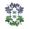
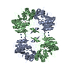
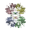
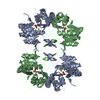
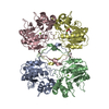
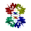
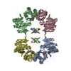
- Assembly
| Deposited unit | 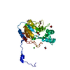
| |||||||||
|---|---|---|---|---|---|---|---|---|---|---|
| 1 | 
| |||||||||
| Unit cell |
| |||||||||
| Components on special symmetry positions |
|
-Components
-Protein , 1 types, 1 molecules A
| #1: Protein | Mass: 23555.342 Da / Num. of mol.: 1 Source method: isolated from a genetically manipulated source Source: (gene. exp.) |
|---|
-Non-polymers , 5 types, 333 molecules 


| #2: Chemical |  Mass: 24.305 Da / Num. of mol.: 2 / Source method: obtained synthetically / Formula: Mg Mass: 24.305 Da / Num. of mol.: 2 / Source method: obtained synthetically / Formula: Mg#3: Chemical | ChemComp-ACT / |  Mass: 59.044 Da / Num. of mol.: 1 / Source method: obtained synthetically / Formula: C2H3O2 Mass: 59.044 Da / Num. of mol.: 1 / Source method: obtained synthetically / Formula: C2H3O2#4: Chemical | ChemComp-CL / |  Mass: 35.453 Da / Num. of mol.: 1 / Source method: obtained synthetically / Formula: Cl Mass: 35.453 Da / Num. of mol.: 1 / Source method: obtained synthetically / Formula: Cl#5: Chemical | ChemComp-EDO /  Mass: 62.068 Da / Num. of mol.: 5 / Source method: obtained synthetically / Formula: C2H6O2 Mass: 62.068 Da / Num. of mol.: 5 / Source method: obtained synthetically / Formula: C2H6O2#6: Water | ChemComp-HOH / | Mass: 18.015 Da / Num. of mol.: 324 / Source method: isolated from a natural source / Formula: H2O |
|---|
-Experimental details
-Experiment
| Experiment | Method: X-RAY DIFFRACTION / Number of used crystals: 1 |
|---|
- Sample preparation
| Crystal | Density Matthews: 3.34 Å3/Da / Density % sol: 63.17 % |
|---|---|
| Crystal grow | Temperature: 279 K / Method: vapor diffusion, sitting drop / pH: 7.2 Details: drop: 4.2-3.5 mg/mL in 50 mM CH3COONa buffer at pH 7.2, 10 mM MgCl2 reservoir: 18% (w/v) polyethylene glycol (PEG) 6000, 0.6% (w/v) Spermine, VAPOR DIFFUSION, SITTING DROP, temperature 279K |
-Data collection
| Diffraction | Mean temperature: 100 K |
|---|---|
| Diffraction source | Source: SYNCHROTRON / Site: ESRF  / Beamline: ID14-4 / Wavelength: 0.934 Å / Beamline: ID14-4 / Wavelength: 0.934 Å |
| Detector | Type: ADSC QUANTUM 4 / Detector: CCD / Date: Jul 13, 2002 Details: Monochromator double crystal, Si(111); toroidal mirror |
| Radiation | Monochromator: double crystal Si(111) / Protocol: SINGLE WAVELENGTH / Monochromatic (M) / Laue (L): M / Scattering type: x-ray |
| Radiation wavelength | Wavelength: 0.934 Å / Relative weight: 1 |
| Reflection | Resolution: 1.7→8 Å / Num. obs: 31967 / % possible obs: 96.8 % / Observed criterion σ(F): 0 / Observed criterion σ(I): 2 / Redundancy: 4.3 % / Biso Wilson estimate: 17.14 Å2 / Rsym value: 0.092 / Net I/σ(I): 4.4 |
| Reflection shell | Resolution: 1.7→1.742 Å / Redundancy: 4.14 % / Rmerge(I) obs: 0.165 / Mean I/σ(I) obs: 2.3 / Num. unique all: 2311 / Rsym value: 0.165 / % possible all: 99.51 |
- Processing
| Software |
| |||||||||||||||||||||||||||||||||||||||||||||||||||||||||||||||||||||||||||||||||||||||||||||||
|---|---|---|---|---|---|---|---|---|---|---|---|---|---|---|---|---|---|---|---|---|---|---|---|---|---|---|---|---|---|---|---|---|---|---|---|---|---|---|---|---|---|---|---|---|---|---|---|---|---|---|---|---|---|---|---|---|---|---|---|---|---|---|---|---|---|---|---|---|---|---|---|---|---|---|---|---|---|---|---|---|---|---|---|---|---|---|---|---|---|---|---|---|---|---|---|---|
| Refinement | Method to determine structure: MOLECULAR REPLACEMENT Starting model: PDB entry 2B82 Resolution: 1.7→7.99 Å / Cor.coef. Fo:Fc: 0.958 / Cor.coef. Fo:Fc free: 0.942 / SU B: 1.728 / SU ML: 0.058 / Isotropic thermal model: isotropic / Cross valid method: THROUGHOUT / σ(F): 0 / σ(I): 2 / ESU R: 0.092 / ESU R Free: 0.093 / Stereochemistry target values: MAXIMUM LIKELIHOOD / Details: HYDROGENS HAVE BEEN ADDED IN THE RIDING POSITIONS
| |||||||||||||||||||||||||||||||||||||||||||||||||||||||||||||||||||||||||||||||||||||||||||||||
| Solvent computation | Ion probe radii: 0.8 Å / Shrinkage radii: 0.8 Å / VDW probe radii: 1.2 Å / Solvent model: MASK | |||||||||||||||||||||||||||||||||||||||||||||||||||||||||||||||||||||||||||||||||||||||||||||||
| Displacement parameters | Biso mean: 23.54 Å2
| |||||||||||||||||||||||||||||||||||||||||||||||||||||||||||||||||||||||||||||||||||||||||||||||
| Refinement step | Cycle: LAST / Resolution: 1.7→7.99 Å
| |||||||||||||||||||||||||||||||||||||||||||||||||||||||||||||||||||||||||||||||||||||||||||||||
| Refine LS restraints |
| |||||||||||||||||||||||||||||||||||||||||||||||||||||||||||||||||||||||||||||||||||||||||||||||
| LS refinement shell | Resolution: 1.7→1.742 Å / Total num. of bins used: 20
|
