 Movie
Movie Controller

[English] 日本語
 Yorodumi
- PDB-2heg: Phospho-Aspartyl Intermediate Analogue of Apha class B acid phosp... -
+ Open data

- Basic information
| Entry | Database: PDB / ID: 2heg | ||||||
|---|---|---|---|---|---|---|---|
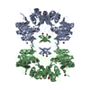
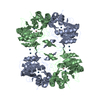
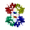
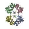
| Title | Phospho-Aspartyl Intermediate Analogue of Apha class B acid phosphatase/phosphotransferase | ||||||
 Components Components | Class B acid phosphatase | ||||||
 Keywords Keywords | HYDROLASE / Apha class B Acid Phosphatase/Phosphotransferase | ||||||
| Function / homology |  Function and homology information Function and homology informationL-phosphoserine phosphatase activity / acid phosphatase / acid phosphatase activity / outer membrane-bounded periplasmic space / magnesium ion binding / identical protein binding Similarity search - Function | ||||||
| Biological species |  | ||||||
| Method |  X-RAY DIFFRACTION / SYNCHROTRON / MOLECULAR REPLACEMENT / Resolution: 1.5 Å X-RAY DIFFRACTION / SYNCHROTRON / MOLECULAR REPLACEMENT / Resolution: 1.5 Å | ||||||
 Authors Authors | Leone, R. / Calderone, V. / Cappelletti, E. / Benvenuti, M. / Mangani, S. | ||||||
 Citation Citation | #1: Journal: J.Mol.Biol. / Year: 2004Title: The First Structure of a Bacterial Class B Acid Phosphatase Reveals Further Structural Heterogeneity Among Phosphatases of Haloacid Deahalogenase Fold Authors: Calderone, V. / Forleo, C. / Benvenuti, M. / Thaller, M.C. / Rossolini, G.M. / Mangani, S. #2: Journal: Biochim.Biophys.Acta / Year: 2006Title: Biochemical Characterization of the Class B Acid Phosphatase (Apha) of Escherichia Coli Mg1655 Authors: Passariello, C. / Forleo, C. / Micheli, V. / Schippa, S. / Leone, R. / Mangani, S. / Thaller, M.C. / Rossolini, G.M. #3: Journal: J.Mol.Biol. / Year: 2006Title: A Structure-Based Proposal for the Catalytic Mechanism of Bacterial Acid Phosphatase Apha Belonging to the Dddd Superfamily of Phosphohydrolases Authors: Calderone, V. / Forleo, C. / Benvenuti, M. / Thaller, M.C. / Rossolini, G.M. / Mangani, S. | ||||||
| History |
|
- Structure visualization
| Structure viewer | Molecule: MolmilJmol/JSmol |
|---|
- Downloads & links
-Download
| PDBx/mmCIF format | 2heg.cif.gz | 116.6 KB | Display | PDBx/mmCIF format |
|---|---|---|---|---|
| PDB format | pdb2heg.ent.gz | 88.3 KB | Display | PDB format |
| PDBx/mmJSON format | 2heg.json.gz | Tree view | PDBx/mmJSON format | |
| Others |  Other downloads Other downloads |
-Validation report
| Arichive directory | https://data.pdbj.org/pub/pdb/validation_reports/he/2hegftp://data.pdbj.org/pub/pdb/validation_reports/he/2heg | HTTPS FTP |
|---|
-Related structure data
| Related structure data |  2b82S S: Starting model for refinement |
|---|---|
| Similar structure data |


-Links
 PDBj
PDBj
- Assembly
| Deposited unit | 
| |||||||||||||||||||||
|---|---|---|---|---|---|---|---|---|---|---|---|---|---|---|---|---|---|---|---|---|---|---|
| 1 | 
| |||||||||||||||||||||
| Unit cell |
| |||||||||||||||||||||
| Components on special symmetry positions |
| |||||||||||||||||||||
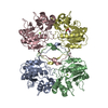
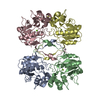
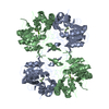
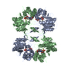
| Details | The biological assembly is a tetramer generated from the dimer in the asymmetric unit by the operations:x,y,z, 1-x,y,1-z |
-Components
| #1: Protein | Mass: 23533.264 Da / Num. of mol.: 2 Source method: isolated from a genetically manipulated source Source: (gene. exp.) #2: Chemical |   Mass: 24.305 Da / Num. of mol.: 2 / Source method: obtained synthetically / Formula: Mg Mass: 24.305 Da / Num. of mol.: 2 / Source method: obtained synthetically / Formula: Mg#3: Water | ChemComp-HOH / |  Mass: 18.015 Da / Num. of mol.: 892 / Source method: isolated from a natural source / Formula: H2O Mass: 18.015 Da / Num. of mol.: 892 / Source method: isolated from a natural source / Formula: H2O |
|---|
-Experimental details
-Experiment
| Experiment | Method: X-RAY DIFFRACTION / Number of used crystals: 1 |
|---|
- Sample preparation
| Crystal | Density Matthews: 2.55 Å3/Da / Density % sol: 51.67 % |
|---|---|
| Crystal grow | Temperature: 279 K / Method: vapor diffusion, sitting drop / pH: 7.2 Details: reservoir: Na acetate 50mM, PEG6000 18% (w/v), 0.6% Spermine (w/v) protein concentration: 3.5 mg/ml, pH 7.2, VAPOR DIFFUSION, SITTING DROP, temperature 279K |
-Data collection
| Diffraction | Mean temperature: 100 K |
|---|---|
| Diffraction source | Source: SYNCHROTRON / Site: ESRF  / Beamline: ID14-4 / Wavelength: 0.939239 Å / Beamline: ID14-4 / Wavelength: 0.939239 Å |
| Detector | Type: ADSC QUANTUM 315 / Detector: CCD / Date: May 17, 2006 |
| Radiation | Monochromator: Khozu monochromator with a McLennon controller containing a LN2 cooled Si111 crystal Protocol: SINGLE WAVELENGTH / Monochromatic (M) / Laue (L): M / Scattering type: x-ray |
| Radiation wavelength | Wavelength: 0.939239 Å / Relative weight: 1 |
| Reflection | Resolution: 1.5→51.23 Å / Num. obs: 75720 / % possible obs: 99.8 % / Redundancy: 7.2 % / Rmerge(I) obs: 0.068 |
| Reflection shell | Resolution: 1.5→1.58 Å / Rmerge(I) obs: 0.358 / % possible all: 100 |
- Processing
| Software |
| |||||||||||||||||||||||||||||||||||||||||||||||||||||||||||||||||||||||||||||||||||||||||||||||
|---|---|---|---|---|---|---|---|---|---|---|---|---|---|---|---|---|---|---|---|---|---|---|---|---|---|---|---|---|---|---|---|---|---|---|---|---|---|---|---|---|---|---|---|---|---|---|---|---|---|---|---|---|---|---|---|---|---|---|---|---|---|---|---|---|---|---|---|---|---|---|---|---|---|---|---|---|---|---|---|---|---|---|---|---|---|---|---|---|---|---|---|---|---|---|---|---|
| Refinement | Method to determine structure: MOLECULAR REPLACEMENT Starting model: PDB code 2B82 Resolution: 1.5→51.23 Å / Cor.coef. Fo:Fc: 0.962 / Cor.coef. Fo:Fc free: 0.952 / Cross valid method: THROUGHOUT / ESU R: 0.072 / ESU R Free: 0.073 / Stereochemistry target values: MAXIMUM LIKELIHOOD / Details: HYDROGENS HAVE BEEN ADDED IN THE RIDING POSITIONS
| |||||||||||||||||||||||||||||||||||||||||||||||||||||||||||||||||||||||||||||||||||||||||||||||
| Solvent computation | Ion probe radii: 0.8 Å / Shrinkage radii: 0.8 Å / VDW probe radii: 1.2 Å / Solvent model: MASK | |||||||||||||||||||||||||||||||||||||||||||||||||||||||||||||||||||||||||||||||||||||||||||||||
| Displacement parameters | Biso mean: 14.772 Å2
| |||||||||||||||||||||||||||||||||||||||||||||||||||||||||||||||||||||||||||||||||||||||||||||||
| Refinement step | Cycle: LAST / Resolution: 1.5→51.23 Å
| |||||||||||||||||||||||||||||||||||||||||||||||||||||||||||||||||||||||||||||||||||||||||||||||
| Refine LS restraints |
| |||||||||||||||||||||||||||||||||||||||||||||||||||||||||||||||||||||||||||||||||||||||||||||||
| LS refinement shell | Resolution: 1.5→1.539 Å / Total num. of bins used: 20
|
