 Movie
Movie Controller

[English] 日本語
 Yorodumi
- PDB-1rmq: Crystal structure of AphA class B acid phosphatase/phosphotransfe... -
+ Open data

- Basic information
| Entry | Database: PDB / ID: 1rmq | ||||||
|---|---|---|---|---|---|---|---|
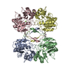
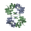
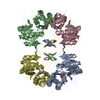
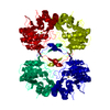
| Title | Crystal structure of AphA class B acid phosphatase/phosphotransferase with osmiate mimicking the catalytic intermediate | ||||||
 Components Components | Class B acid phosphatase | ||||||
 Keywords Keywords | HYDROLASE / Class B acid phosphatase / DDDD acid phosphatase / metallo-enzyme / osmiate | ||||||
| Function / homology |  Function and homology information Function and homology informationL-phosphoserine phosphatase activity / acid phosphatase / acid phosphatase activity / outer membrane-bounded periplasmic space / magnesium ion binding / metal ion binding / identical protein binding Similarity search - Function | ||||||
| Biological species |  | ||||||
| Method |  X-RAY DIFFRACTION / SYNCHROTRON / MOLECULAR REPLACEMENT / Resolution: 2 Å X-RAY DIFFRACTION / SYNCHROTRON / MOLECULAR REPLACEMENT / Resolution: 2 Å | ||||||
 Authors Authors | Calderone, V. / Forleo, C. / Benvenuti, M. / Rossolini, G.M. / Thaller, M.C. / Mangani, S. | ||||||
 Citation Citation | Journal: To be Published Title: Insights in the catalytic mechanism of AphA from Escherichia coli Authors: Calderone, V. / Forleo, C. / Benvenuti, M. / Rossolini, G.M. / Thaller, M.C. / Mangani, S. #1: Journal: FEMS Microbiol.Lett. / Year: 1997Title: Identification of the gene (aphA) encoding the class B acid phosphatase/phosphotransferase of Escherichia coli MG1655 and characterization of its product Authors: Thaller, M.C. / Schippa, S. / Bonci, A. / Cresti, S. / Rossolini, G.M. #2: Journal: Acta Crystallogr.,Sect.D / Year: 2003Title: Expression, purification, crystallization and preliminary X-ray characterization of the class B acid phosphatase (AphA) from Escherichia coli Authors: Forleo, C. / Benvenuti, M. / Calderone, V. / Schippa, S. / Docquier, J.D. / Thaller, M.C. / M Rossolini, G. / Mangani, S. | ||||||
| History |
|
- Structure visualization
| Structure viewer | Molecule: MolmilJmol/JSmol |
|---|
- Downloads & links
-Download
| PDBx/mmCIF format | 1rmq.cif.gz | 102.5 KB | Display | PDBx/mmCIF format |
|---|---|---|---|---|
| PDB format | pdb1rmq.ent.gz | 78.1 KB | Display | PDB format |
| PDBx/mmJSON format | 1rmq.json.gz | Tree view | PDBx/mmJSON format | |
| Others |  Other downloads Other downloads |
-Validation report
| Arichive directory | https://data.pdbj.org/pub/pdb/validation_reports/rm/1rmqftp://data.pdbj.org/pub/pdb/validation_reports/rm/1rmq | HTTPS FTP |
|---|
-Related structure data
| Related structure data |  1rmtC  1rmyC  1n8nS S: Starting model for refinement C: citing same article ( |
|---|---|
| Similar structure data |


-Links
 PDBj
PDBj

- Assembly
| Deposited unit | 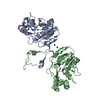
| |||||||||
|---|---|---|---|---|---|---|---|---|---|---|
| 1 | 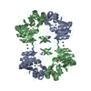
| |||||||||
| Unit cell |
| |||||||||
| Components on special symmetry positions |
| |||||||||
| Details | The biological assembly is a tetramer, but there is a dimer in the asymmetric unit. |
-Components
| #1: Protein | Mass: 23555.342 Da / Num. of mol.: 2 Source method: isolated from a genetically manipulated source Source: (gene. exp.) References: UniProt: P32697, UniProt: P0AE22*PLUS, acid phosphatase #2: Chemical |   Mass: 58.933 Da / Num. of mol.: 2 / Source method: obtained synthetically / Formula: Co Mass: 58.933 Da / Num. of mol.: 2 / Source method: obtained synthetically / Formula: Co#3: Chemical | ChemComp-OS /   Mass: 190.230 Da / Num. of mol.: 4 / Source method: obtained synthetically / Formula: Os Mass: 190.230 Da / Num. of mol.: 4 / Source method: obtained synthetically / Formula: Os#4: Water | ChemComp-HOH / |  Mass: 18.015 Da / Num. of mol.: 296 / Source method: isolated from a natural source / Formula: H2O Mass: 18.015 Da / Num. of mol.: 296 / Source method: isolated from a natural source / Formula: H2O |
|---|
-Experimental details
-Experiment
| Experiment | Method: X-RAY DIFFRACTION / Number of used crystals: 1 |
|---|
- Sample preparation
| Crystal | Density Matthews: 2.59 Å3/Da / Density % sol: 52.11 % |
|---|---|
| Crystal grow | Temperature: 293 K / Method: vapor diffusion, sitting drop / pH: 7.2 Details: AphA 6mg/mL, 50mM Na acetate, 25% PEG 6000, 10mM osmiate, pH 7.2, VAPOR DIFFUSION, SITTING DROP, temperature 293K |
-Data collection
| Diffraction | Mean temperature: 100 K | ||||||||||||
|---|---|---|---|---|---|---|---|---|---|---|---|---|---|
| Diffraction source | Source: SYNCHROTRON / Site: ESRF  / Beamline: BM30A / Wavelength: 1.13980, 1.14057, 1.13800 / Beamline: BM30A / Wavelength: 1.13980, 1.14057, 1.13800 | ||||||||||||
| Detector | Type: MARRESEARCH / Detector: CCD / Date: Feb 12, 2002 / Details: Double crystal monochromator | ||||||||||||
| Radiation | Monochromator: Double crystal monochromator / Protocol: MAD / Monochromatic (M) / Laue (L): M / Scattering type: x-ray | ||||||||||||
| Radiation wavelength |
| ||||||||||||
| Reflection | Resolution: 2→30.58 Å / Num. all: 31710 / Num. obs: 31710 / % possible obs: 99.3 % / Observed criterion σ(F): 0 / Observed criterion σ(I): 0 / Redundancy: 3.4 % / Biso Wilson estimate: 23.344 Å2 / Rmerge(I) obs: 0.089 / Rsym value: 0.089 / Net I/σ(I): 6.2 | ||||||||||||
| Reflection shell | Resolution: 2→2.11 Å / Redundancy: 3.1 % / Rmerge(I) obs: 0.402 / Mean I/σ(I) obs: 1.6 / Num. unique all: 4531 / Rsym value: 0.402 / % possible all: 97.6 |
- Processing
| Software |
| |||||||||||||||||||||||||||||||||||||||||||||||||||||||||||||||||||||||||||
|---|---|---|---|---|---|---|---|---|---|---|---|---|---|---|---|---|---|---|---|---|---|---|---|---|---|---|---|---|---|---|---|---|---|---|---|---|---|---|---|---|---|---|---|---|---|---|---|---|---|---|---|---|---|---|---|---|---|---|---|---|---|---|---|---|---|---|---|---|---|---|---|---|---|---|---|---|
| Refinement | Method to determine structure: MOLECULAR REPLACEMENT Starting model: 1N8N Resolution: 2→30 Å / Cor.coef. Fo:Fc: 0.942 / Cor.coef. Fo:Fc free: 0.921 / SU B: 4.04 / SU ML: 0.113 / Cross valid method: THROUGHOUT / σ(F): 0 / σ(I): 0 / ESU R: 0.183 / ESU R Free: 0.165 / Stereochemistry target values: MAXIMUM LIKELIHOOD
| |||||||||||||||||||||||||||||||||||||||||||||||||||||||||||||||||||||||||||
| Solvent computation | Ion probe radii: 0.8 Å / Shrinkage radii: 0.8 Å / VDW probe radii: 1.4 Å / Solvent model: BABINET MODEL WITH MASK | |||||||||||||||||||||||||||||||||||||||||||||||||||||||||||||||||||||||||||
| Displacement parameters | Biso mean: 26.179 Å2
| |||||||||||||||||||||||||||||||||||||||||||||||||||||||||||||||||||||||||||
| Refine analyze | Luzzati coordinate error obs: 0.113 Å | |||||||||||||||||||||||||||||||||||||||||||||||||||||||||||||||||||||||||||
| Refinement step | Cycle: LAST / Resolution: 2→30 Å
| |||||||||||||||||||||||||||||||||||||||||||||||||||||||||||||||||||||||||||
| Refine LS restraints |
| |||||||||||||||||||||||||||||||||||||||||||||||||||||||||||||||||||||||||||
| LS refinement shell | Resolution: 2→2.052 Å / Total num. of bins used: 20 /
|
