 Movie
Movie Controller

[English] 日本語
 Yorodumi
- PDB-2b82: Crystal structure of AphA class B acid phosphatase/phosphotransfe... -
+ Open data

- Basic information
| Entry | Database: PDB / ID: 2b82 | |||||||||
|---|---|---|---|---|---|---|---|---|---|---|
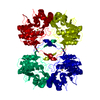
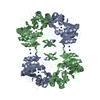
| Title | Crystal structure of AphA class B acid phosphatase/phosphotransferase ternary complex with adenosine and phosphate bound to the catalytic metal at 1.2 A resolution | |||||||||
 Components Components | class B acid phosphatase | |||||||||
 Keywords Keywords | HYDROLASE / Class B acid phosphatase / DDDD acid phosphatase / metallo-enzyme / AMP | |||||||||
| Function / homology |  Function and homology information Function and homology informationL-phosphoserine phosphatase activity / acid phosphatase / acid phosphatase activity / outer membrane-bounded periplasmic space / magnesium ion binding / metal ion binding / identical protein binding Similarity search - Function | |||||||||
| Biological species |  | |||||||||
| Method |  X-RAY DIFFRACTION / SYNCHROTRON / MOLECULAR REPLACEMENT / Resolution: 1.25 Å X-RAY DIFFRACTION / SYNCHROTRON / MOLECULAR REPLACEMENT / Resolution: 1.25 Å | |||||||||
 Authors Authors | Calderone, V. / Forleo, C. / Benvenuti, M. / Thaller, M.C. / Rossolini, G.M. / Mangani, S. | |||||||||
 Citation Citation | Journal: J.Mol.Biol. / Year: 2006 Title: A structure-based proposal for the catalytic mechanism of the bacterial acid phosphatase AphA belonging to the DDDD superfamily of phosphohydrolases Authors: Calderone, V. / Forleo, C. / Benvenuti, M. / Thaller, M.C. / Rossolini, G.M. / Mangani, S. #1: Journal: FEMS Microbiol.Lett. / Year: 1997Title: Identification of the gene (aphA) encoding the class B acid phosphatase/phosphotransferase of Escherichia coli MG1655 and characterization of its product Authors: Thaller, M.C. / Schippa, S. / Bonci, A. / Cresti, S. / Rossolini, G.M. #2: Journal: Acta Crystallogr.,Sect.D / Year: 2003Title: Expression, purification, crystallization and preliminary X-ray characterization of the class B acid phosphatase (AphA) from Escherichia coli Authors: Forleo, C. / Benvenuti, M. / Calderone, V. / Schippa, S. / Docquier, J.D. / Thaller, M.C. / Rossolini, G.M. / Mangani, S. | |||||||||
| History |
|
- Structure visualization
| Structure viewer | Molecule: MolmilJmol/JSmol |
|---|
- Downloads & links
-Download
| PDBx/mmCIF format | 2b82.cif.gz | 209.3 KB | Display | PDBx/mmCIF format |
|---|---|---|---|---|
| PDB format | pdb2b82.ent.gz | 164.8 KB | Display | PDB format |
| PDBx/mmJSON format | 2b82.json.gz | Tree view | PDBx/mmJSON format | |
| Others |  Other downloads Other downloads |
-Validation report
| Arichive directory | https://data.pdbj.org/pub/pdb/validation_reports/b8/2b82ftp://data.pdbj.org/pub/pdb/validation_reports/b8/2b82 | HTTPS FTP |
|---|
-Related structure data
| Related structure data |  2b8jC  1n8nS C: citing same article ( S: Starting model for refinement |
|---|---|
| Similar structure data |


-Links
 PDBj
PDBj

- Assembly
| Deposited unit | 
| |||||||||||||||
|---|---|---|---|---|---|---|---|---|---|---|---|---|---|---|---|---|
| 1 | 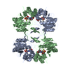
| |||||||||||||||
| Unit cell |
| |||||||||||||||
| Components on special symmetry positions |
| |||||||||||||||
| Details | The biological assembly is a tetramer, but there is a dimer in the asymmetric unit |
-Components
| #1: Protein | Mass: 23468.264 Da / Num. of mol.: 2 Source method: isolated from a genetically manipulated source Source: (gene. exp.) References: UniProt: P32697, UniProt: P0AE22*PLUS, acid phosphatase #2: Chemical |   Mass: 24.305 Da / Num. of mol.: 2 / Source method: obtained synthetically / Formula: Mg Mass: 24.305 Da / Num. of mol.: 2 / Source method: obtained synthetically / Formula: Mg#3: Chemical |   Mass: 94.971 Da / Num. of mol.: 2 / Source method: obtained synthetically / Formula: PO4 Mass: 94.971 Da / Num. of mol.: 2 / Source method: obtained synthetically / Formula: PO4#4: Chemical |   Mass: 267.241 Da / Num. of mol.: 2 / Source method: obtained synthetically / Formula: C10H13N5O4 Mass: 267.241 Da / Num. of mol.: 2 / Source method: obtained synthetically / Formula: C10H13N5O4#5: Water | ChemComp-HOH / |  Mass: 18.015 Da / Num. of mol.: 856 / Source method: isolated from a natural source / Formula: H2O Mass: 18.015 Da / Num. of mol.: 856 / Source method: isolated from a natural source / Formula: H2O |
|---|
-Experimental details
-Experiment
| Experiment | Method: X-RAY DIFFRACTION / Number of used crystals: 1 |
|---|
- Sample preparation
| Crystal | Density Matthews: 2.4 Å3/Da / Density % sol: 48.9 % |
|---|---|
| Crystal grow | Temperature: 293 K / Method: vapor diffusion, sitting drop / pH: 7.2 Details: AphA 6mg/mL, 50 mM Na acetate, 25% PEG 6000, 10mM AMP, pH 7.2, VAPOR DIFFUSION, SITTING DROP, temperature 293K |
-Data collection
| Diffraction | Mean temperature: 100 K |
|---|---|
| Diffraction source | Source: SYNCHROTRON / Site: ESRF  / Beamline: ID29 / Wavelength: 0.96111 Å / Beamline: ID29 / Wavelength: 0.96111 Å |
| Detector | Type: ADSC QUANTUM 4 / Detector: CCD / Date: Dec 7, 2002 Details: channel - cut Si monochromator and cylindrical grazing incidence mirror |
| Radiation | Monochromator: Si (311) and Si (111) / Protocol: SINGLE WAVELENGTH / Monochromatic (M) / Laue (L): M / Scattering type: x-ray |
| Radiation wavelength | Wavelength: 0.96111 Å / Relative weight: 1 |
| Reflection | Resolution: 1.25→76.71 Å / Num. all: 112327 / Num. obs: 112327 / % possible obs: 88.7 % / Observed criterion σ(F): 0 / Observed criterion σ(I): 0 / Redundancy: 2.6 % / Biso Wilson estimate: 8.827 Å2 / Rmerge(I) obs: 0.069 / Rsym value: 0.069 / Net I/σ(I): 5.1 |
| Reflection shell | Resolution: 1.25→1.32 Å / Redundancy: 1.8 % / Rmerge(I) obs: 0.22 / Mean I/σ(I) obs: 3 / Num. unique all: 10703 / Rsym value: 0.22 / % possible all: 58.2 |
- Processing
| Software |
| ||||||||||||||||||||||||||||||||||||||||||||||||||||||||||||||||||||||||||||||||||||||||||||||||||||||||||||||||||||||||||||||||||||||||||||||||||||||||||||||||||||||||||
|---|---|---|---|---|---|---|---|---|---|---|---|---|---|---|---|---|---|---|---|---|---|---|---|---|---|---|---|---|---|---|---|---|---|---|---|---|---|---|---|---|---|---|---|---|---|---|---|---|---|---|---|---|---|---|---|---|---|---|---|---|---|---|---|---|---|---|---|---|---|---|---|---|---|---|---|---|---|---|---|---|---|---|---|---|---|---|---|---|---|---|---|---|---|---|---|---|---|---|---|---|---|---|---|---|---|---|---|---|---|---|---|---|---|---|---|---|---|---|---|---|---|---|---|---|---|---|---|---|---|---|---|---|---|---|---|---|---|---|---|---|---|---|---|---|---|---|---|---|---|---|---|---|---|---|---|---|---|---|---|---|---|---|---|---|---|---|---|---|---|---|---|
| Refinement | Method to determine structure: MOLECULAR REPLACEMENT Starting model: 1N8N Resolution: 1.25→76.71 Å / Cor.coef. Fo:Fc: 0.963 / Cor.coef. Fo:Fc free: 0.954 / SU B: 1.256 / SU ML: 0.026 / Isotropic thermal model: anisotropic / Cross valid method: THROUGHOUT / σ(F): 0 / σ(I): 0 / ESU R: 0.054 / ESU R Free: 0.049 / Stereochemistry target values: MAXIMUM LIKELIHOOD / Details: HYDROGENS HAVE BEEN ADDED IN THE RIDING POSITIONS
| ||||||||||||||||||||||||||||||||||||||||||||||||||||||||||||||||||||||||||||||||||||||||||||||||||||||||||||||||||||||||||||||||||||||||||||||||||||||||||||||||||||||||||
| Solvent computation | Ion probe radii: 0.8 Å / Shrinkage radii: 0.8 Å / VDW probe radii: 1.2 Å / Solvent model: MASK | ||||||||||||||||||||||||||||||||||||||||||||||||||||||||||||||||||||||||||||||||||||||||||||||||||||||||||||||||||||||||||||||||||||||||||||||||||||||||||||||||||||||||||
| Displacement parameters | Biso mean: 13.61 Å2
| ||||||||||||||||||||||||||||||||||||||||||||||||||||||||||||||||||||||||||||||||||||||||||||||||||||||||||||||||||||||||||||||||||||||||||||||||||||||||||||||||||||||||||
| Refine analyze | Luzzati coordinate error obs: 0.026 Å | ||||||||||||||||||||||||||||||||||||||||||||||||||||||||||||||||||||||||||||||||||||||||||||||||||||||||||||||||||||||||||||||||||||||||||||||||||||||||||||||||||||||||||
| Refinement step | Cycle: LAST / Resolution: 1.25→76.71 Å
| ||||||||||||||||||||||||||||||||||||||||||||||||||||||||||||||||||||||||||||||||||||||||||||||||||||||||||||||||||||||||||||||||||||||||||||||||||||||||||||||||||||||||||
| Refine LS restraints |
| ||||||||||||||||||||||||||||||||||||||||||||||||||||||||||||||||||||||||||||||||||||||||||||||||||||||||||||||||||||||||||||||||||||||||||||||||||||||||||||||||||||||||||
| LS refinement shell | Resolution: 1.25→1.282 Å / Total num. of bins used: 20
|
