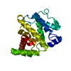
RESIDUES A161-A167 THAT ARE PRESENT IN COORDINATES ARE LIKELY PART OF ENTITY 1. THEY WERE MODELED ...RESIDUES A161-A167 THAT ARE PRESENT IN COORDINATES ARE LIKELY PART OF ENTITY 1. THEY WERE MODELED AS POLY-ALA. THEIR ASSIGNMENT TO THE AMINO ACID SEQUENCE OF THE PROTEIN IS UNKNOWN DUE TO LACK OF CONTINUOUS ELECTRON DENSITY.
解像度: 2.6→28.513 Å / Cor.coef. Fo:Fc: 0.895 / Cor.coef. Fo:Fc free: 0.863 / WRfactor Rfree: 0.298 / WRfactor Rwork: 0.263 / SU B: 25.513 / SU ML: 0.262 / TLS residual ADP flag: LIKELY RESIDUAL / 交差検証法: THROUGHOUT / σ(F): 0 / ESU R: 0.333 / ESU R Free: 0.273 / 立体化学のターゲット値: MAXIMUM LIKELIHOOD 詳細: 1. HYDROGENS HAVE BEEN ADDED IN THE RIDING POSITIONS. 2. Atomic B-factors are residuals from TLS refinement. 3. Programs coot, molprobity, ffas03, SCWRL have also been used in refinement. 4. ...詳細: 1. HYDROGENS HAVE BEEN ADDED IN THE RIDING POSITIONS. 2. Atomic B-factors are residuals from TLS refinement. 3. Programs coot, molprobity, ffas03, SCWRL have also been used in refinement. 4. Residues A 161 through A 167 are likely part of entity 1. They were modeled as poly-Ala due to insufficient electron density. Their assignment to the amino acid sequence is unknown due to lack of continuous electron density.
Rfactor
反射数
%反射
Selection details
Rfree
0.3
1433
5.062 %
RANDOM
Rwork
0.266
-
-
-
obs
0.267
28308
99.546 %
-
溶媒の処理
イオンプローブ半径: 0.8 Å / 減衰半径: 0.8 Å / VDWプローブ半径: 1.4 Å / 溶媒モデル: MASK BULK SOLVENT
原子変位パラメータ
Biso mean: 48.663 Å2
Baniso -1
Baniso -2
Baniso -3
1-
0.044 Å2
0.022 Å2
0 Å2
2-
-
0.044 Å2
0 Å2
3-
-
-
-0.067 Å2
精密化ステップ
サイクル: LAST / 解像度: 2.6→28.513 Å
タンパク質
核酸
リガンド
溶媒
全体
原子数
3167
0
3
0
3170
拘束条件
Refine-ID
タイプ
Dev ideal
Dev ideal target
数
X-RAY DIFFRACTION
r_bond_refined_d
0.012
0.022
3245
X-RAY DIFFRACTION
r_bond_other_d
0.002
0.02
2184
X-RAY DIFFRACTION
r_angle_refined_deg
1.053
1.963
4392
X-RAY DIFFRACTION
r_angle_other_deg
0.815
3.002
5306
X-RAY DIFFRACTION
r_dihedral_angle_1_deg
5.211
5
399
X-RAY DIFFRACTION
r_dihedral_angle_2_deg
31.79
23.537
147
X-RAY DIFFRACTION
r_dihedral_angle_3_deg
15.03
15
547
X-RAY DIFFRACTION
r_dihedral_angle_4_deg
21.059
15
21
X-RAY DIFFRACTION
r_chiral_restr
0.057
0.2
488
X-RAY DIFFRACTION
r_gen_planes_refined
0.003
0.02
3597
X-RAY DIFFRACTION
r_gen_planes_other
0.001
0.02
667
X-RAY DIFFRACTION
r_nbd_refined
0.218
0.2
709
X-RAY DIFFRACTION
r_nbd_other
0.173
0.2
2152
X-RAY DIFFRACTION
r_nbtor_refined
0.179
0.2
1577
X-RAY DIFFRACTION
r_nbtor_other
0.082
0.2
1579
X-RAY DIFFRACTION
r_xyhbond_nbd_refined
0.133
0.2
57
X-RAY DIFFRACTION
r_xyhbond_nbd_other
0.001
0.2
1
X-RAY DIFFRACTION
r_symmetry_vdw_refined
0.197
0.2
11
X-RAY DIFFRACTION
r_symmetry_vdw_other
0.227
0.2
18
X-RAY DIFFRACTION
r_symmetry_hbond_refined
0.063
0.2
1
X-RAY DIFFRACTION
r_mcbond_it
0.342
1.5
2066
X-RAY DIFFRACTION
r_mcbond_other
0.052
1.5
806
X-RAY DIFFRACTION
r_mcangle_it
0.576
2
3215
X-RAY DIFFRACTION
r_scbond_it
0.792
3
1346
X-RAY DIFFRACTION
r_scangle_it
1.241
4.5
1177
LS精密化 シェル
Refine-ID: X-RAY DIFFRACTION / Total num. of bins used: 20
 ムービー
ムービー コントローラー


 データを開く
 基本情報
基本情報 要素
要素 キーワード
キーワード 機能・相同性情報
 Homo sapiens (ヒト)
 X線回折 /
  データ登録者
 引用
引用 構造の表示
構造の表示 ダウンロードとリンク
 その他のダウンロード


 PDBj
PDBj


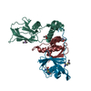
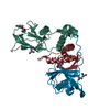
 集合体
集合体


 分子量: 65.409 Da / 分子数: 2 / 由来タイプ: 合成 / 式: Zn
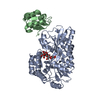
 分子数: 1 / 由来タイプ: 合成
 試料調製
試料調製 / ビームライン: 19-ID / 波長: 0.99987 Å
 解析
解析