 Movie
Movie Controller

[English] 日本語
 Yorodumi
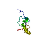
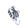
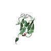
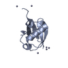
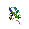
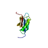
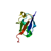
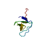
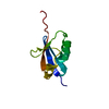
- PDB-3cwi: Crystal structure of thiamine biosynthesis protein (ThiS) from Ge... -
+ Open data

- Basic information
| Entry | Database: PDB / ID: 3cwi | ||||||
|---|---|---|---|---|---|---|---|
| Title | Crystal structure of thiamine biosynthesis protein (ThiS) from Geobacter metallireducens. Northeast Structural Genomics Consortium Target GmR137 | ||||||
 Components Components | Thiamine-biosynthesis protein ThiS | ||||||
 Keywords Keywords | BIOSYNTHETIC PROTEIN / alpha-beta protein / Structural Genomics / PSI-2 / Protein Structure Initiative / Northeast Structural Genomics Consortium / NESG | ||||||
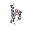
| Function / homology |  Function and homology information Function and homology informationThiS, thiamine-biosynthesis / Sulfur carrier ThiS/MoaD-like / ThiS family / Molybdopterin synthase/thiamin biosynthesis sulphur carrier, beta-grasp / Beta-grasp domain / Beta-grasp domain superfamily / Ubiquitin-like (UB roll) / Roll / Alpha Beta Similarity search - Domain/homology | ||||||
| Biological species |  Geobacter metallireducens GS-15 (bacteria) Geobacter metallireducens GS-15 (bacteria) | ||||||
| Method |  X-RAY DIFFRACTION / SYNCHROTRON / SAD / Resolution: 1.9 Å X-RAY DIFFRACTION / SYNCHROTRON / SAD / Resolution: 1.9 Å | ||||||
 Authors Authors | Forouhar, F. / Abashidze, M. / Seetharaman, J. / Mao, L. / Janjua, H. / Xiao, R. / Maglaqui, M. / Ciccosanti, C. / Foote, E.L. / Wang, H. ...Forouhar, F. / Abashidze, M. / Seetharaman, J. / Mao, L. / Janjua, H. / Xiao, R. / Maglaqui, M. / Ciccosanti, C. / Foote, E.L. / Wang, H. / Everett, J.K. / Acton, T.B. / Montelione, G.T. / Tong, L. / Hunt, J.F. / Northeast Structural Genomics Consortium (NESG) | ||||||
 Citation Citation | Journal: To be Published Title: Crystal structure of thiamine biosynthesis protein (ThiS) from Geobacter metallireducens. Authors: Forouhar, F. / Abashidze, M. / Seetharaman, J. / Mao, L. / Janjua, H. / Xiao, R. / Maglaqui, M. / Ciccosanti, C. / Foote, E.L. / Wang, H. / Everett, J.K. / Acton, T.B. / Montelione, G.T. / Tong, L. / Hunt, J.F. | ||||||
| History |
|
- Structure visualization
| Structure viewer | Molecule: MolmilJmol/JSmol |
|---|
- Downloads & links
-Download
| PDBx/mmCIF format | 3cwi.cif.gz | 26 KB | Display | PDBx/mmCIF format |
|---|---|---|---|---|
| PDB format | pdb3cwi.ent.gz | 16.5 KB | Display | PDB format |
| PDBx/mmJSON format | 3cwi.json.gz | Tree view | PDBx/mmJSON format | |
| Others |  Other downloads Other downloads |
-Validation report
| Arichive directory | https://data.pdbj.org/pub/pdb/validation_reports/cw/3cwiftp://data.pdbj.org/pub/pdb/validation_reports/cw/3cwi | HTTPS FTP |
|---|
-Related structure data
| Similar structure data | |
|---|---|
| Other databases |
-Links
 PDBj
PDBj


- Assembly
| Deposited unit | 
| ||||||||
|---|---|---|---|---|---|---|---|---|---|
| 1 |
| ||||||||
| Unit cell |
| ||||||||
| Details | AUTHORS STATE THAT THE MONOMERIC ASSEMBLY OF THE BIOLOGICAL UNIT THAT IS SHOWN IN REMARK 350 IS PUTATIVE |
-Components
| #1: Protein | Mass: 8624.253 Da / Num. of mol.: 1 Source method: isolated from a genetically manipulated source Source: (gene. exp.) Geobacter metallireducens GS-15 (bacteria)Species: Geobacter metallireducens / Strain: GS-15 / DSM 7210 / Gene: ThiS, Gmet_1567 / Plasmid: pET21 / Production host: |
|---|---|
| #2: Water | ChemComp-HOH /  Mass: 18.015 Da / Num. of mol.: 45 / Source method: isolated from a natural source / Formula: H2O Mass: 18.015 Da / Num. of mol.: 45 / Source method: isolated from a natural source / Formula: H2O |
| Has protein modification | Y |
-Experimental details
-Experiment
| Experiment | Method: X-RAY DIFFRACTION / Number of used crystals: 1 |
|---|
- Sample preparation
| Crystal | Density Matthews: 2.18 Å3/Da / Density % sol: 43.52 % Description: The structure factor file contains Friedel pairs |
|---|---|
| Crystal grow | Temperature: 291 K / Method: vapor diffusion, hanging drop / pH: 5 Details: Protein solution: 10 mM Tris-HCl pH 7.5, 100 mM NaCl, 5 mM DTT. Reservoir solution: 100 mM Sodium acetate pH 5.0, 18% PEG 8000, 200 mM Magnesium chloride, VAPOR DIFFUSION, HANGING DROP, temperature 291K |
-Data collection
| Diffraction | Mean temperature: 100 K |
|---|---|
| Diffraction source | Source: SYNCHROTRON / Site: NSLS  / Beamline: X4C / Wavelength: 0.97908 Å / Beamline: X4C / Wavelength: 0.97908 Å |
| Detector | Type: MAR CCD 165 mm / Detector: CCD / Date: Apr 2, 2008 / Details: Mirrors |
| Radiation | Monochromator: Si 111 CHANNEL / Protocol: SINGLE WAVELENGTH / Monochromatic (M) / Laue (L): M / Scattering type: x-ray |
| Radiation wavelength | Wavelength: 0.97908 Å / Relative weight: 1 |
| Reflection | Resolution: 1.9→30 Å / Num. all: 11436 / Num. obs: 11436 / % possible obs: 99.5 % / Observed criterion σ(F): 0 / Observed criterion σ(I): 0 / Redundancy: 14.5 % / Biso Wilson estimate: 16 Å2 / Rmerge(I) obs: 0.078 / Rsym value: 0.063 / Net I/σ(I): 40.74 |
| Reflection shell | Resolution: 1.9→1.97 Å / Redundancy: 10.8 % / Rmerge(I) obs: 0.37 / Mean I/σ(I) obs: 4.54 / Num. unique all: 1152 / Rsym value: 0.328 / % possible all: 99.8 |
- Processing
| Software |
| |||||||||||||||||||||||||
|---|---|---|---|---|---|---|---|---|---|---|---|---|---|---|---|---|---|---|---|---|---|---|---|---|---|---|
| Refinement | Method to determine structure: SAD / Resolution: 1.9→19.97 Å / Rfactor Rfree error: 0.008 / Data cutoff high absF: 856078.58 / Data cutoff low absF: 0 / Isotropic thermal model: OVERALL / Cross valid method: THROUGHOUT / σ(F): 2 / Stereochemistry target values: Engh & Huber / Details: The Friedel pairs were used in phasing
| |||||||||||||||||||||||||
| Solvent computation | Solvent model: FLAT MODEL / Bsol: 59.014 Å2 / ksol: 0.45 e/Å3 | |||||||||||||||||||||||||
| Displacement parameters | Biso mean: 32 Å2
| |||||||||||||||||||||||||
| Refine analyze |
| |||||||||||||||||||||||||
| Refinement step | Cycle: LAST / Resolution: 1.9→19.97 Å
| |||||||||||||||||||||||||
| Refine LS restraints |
| |||||||||||||||||||||||||
| LS refinement shell | Resolution: 1.9→1.97 Å / Rfactor Rfree error: 0.025 / Total num. of bins used: 10
|
