Type: MARMOSAIC 325 mm CCD / Detector: CCD / Date: Feb 4, 2008 / Details: Flat mirror (vertical focusing)
Radiation
Monochromator: Single crystal Si(111) bent (horizontal focusing) Protocol: MAD / Monochromatic (M) / Laue (L): M / Scattering type: x-ray
Radiation wavelength
ID
Wavelength (Å)
Relative weight
1
0.92522
1
2
0.97922
1
3
0.97464
1
Reflection
Resolution: 2.09→26.528 Å / Num. obs: 9659 / % possible obs: 97.6 % / Observed criterion σ(I): -3 / Biso Wilson estimate: 44.118 Å2 / Rmerge(I) obs: 0.052 / Net I/σ(I): 15.3
Reflection shell
Resolution (Å)
Rmerge(I) obs
Mean I/σ(I) obs
Num. measured obs
Num. unique obs
Diffraction-ID
% possible all
2.09-2.16
0.902
1.6
4806
1296
1
77.8
2.16-2.25
0.67
2.2
7068
1841
1
99.6
2.25-2.35
0.522
2.8
6573
1718
1
99.5
2.35-2.48
0.357
4
7064
1836
1
99.6
2.48-2.63
0.218
6.2
6598
1713
1
99.9
2.63-2.83
0.138
9.5
6746
1751
1
99.9
2.83-3.12
0.077
15.5
6986
1804
1
99.9
3.12-3.57
0.042
25.3
6817
1761
1
99.9
3.57-4.49
0.028
37.1
6793
1766
1
99.9
4.49-26.528
0.023
45
6874
1793
1
98.8
-
Phasing
Phasing
Method: MAD
-
Processing
Software
Name
Version
Classification
NB
REFMAC
5.4.0067
refinement
PHENIX
refinement
SHELX
phasing
MolProbity
3beta29
modelbuilding
XSCALE
datascaling
PDB_EXTRACT
3.004
dataextraction
MAR345
CCD
datacollection
XDS
datareduction
SHARP
phasing
SHELXD
phasing
Refinement
Method to determine structure: MAD / Resolution: 2.1→26.528 Å / Cor.coef. Fo:Fc: 0.952 / Cor.coef. Fo:Fc free: 0.946 / SU B: 12.209 / SU ML: 0.165 / TLS residual ADP flag: LIKELY RESIDUAL / Cross valid method: THROUGHOUT / σ(F): 0 / ESU R: 0.233 / ESU R Free: 0.187 Stereochemistry target values: MAXIMUM LIKELIHOOD WITH PHASES Details: 1. HYDROGENS HAVE BEEN ADDED IN THE RIDING POSITIONS. 2. ATOM RECORDS CONTAIN RESIDUAL B FACTORS ONLY. 3. A MET-INHIBITION PROTOCOL WAS USED FOR SELENOMETHIONINE INCORPORATION DURING PROTEIN ...Details: 1. HYDROGENS HAVE BEEN ADDED IN THE RIDING POSITIONS. 2. ATOM RECORDS CONTAIN RESIDUAL B FACTORS ONLY. 3. A MET-INHIBITION PROTOCOL WAS USED FOR SELENOMETHIONINE INCORPORATION DURING PROTEIN EXPRESSION. THE OCCUPANCY OF THE SE ATOMS IN THE MSE RESIDUES WAS REDUCED TO 0.75 FOR THE REDUCED SCATTERING POWER DUE TO PARTIAL S-MET INCORPORATION. 4. X-RAY FLUORESCENCE EXCITATION, WAVELENGTH SCANS AND ANOMALOUS DIFFERENCE FOURIERS SUPPORT THE MODELING OF ZN ION. 5. AN UNKNOWN LIGAND (UNL) HAS BEEN MODELED NEAR THE ZN ION SITE.
Rfactor
Num. reflection
% reflection
Selection details
Rfree
0.246
461
4.8 %
RANDOM
Rwork
0.216
-
-
-
obs
0.217
9609
99.59 %
-
Solvent computation
Ion probe radii: 0.8 Å / Shrinkage radii: 0.8 Å / VDW probe radii: 1.2 Å / Solvent model: BABINET MODEL WITH MASK
Displacement parameters
Biso mean: 46.793 Å2
Baniso -1
Baniso -2
Baniso -3
1-
0.91 Å2
0 Å2
0 Å2
2-
-
0.91 Å2
0 Å2
3-
-
-
-1.82 Å2
Refinement step
Cycle: LAST / Resolution: 2.1→26.528 Å
Protein
Nucleic acid
Ligand
Solvent
Total
Num. atoms
1093
0
27
28
1148
Refine LS restraints
Refine-ID
Type
Dev ideal
Dev ideal target
Number
X-RAY DIFFRACTION
r_bond_refined_d
0.015
0.021
1158
X-RAY DIFFRACTION
r_bond_other_d
0.003
0.02
806
X-RAY DIFFRACTION
r_angle_refined_deg
1.918
1.947
1570
X-RAY DIFFRACTION
r_angle_other_deg
1.558
3
1951
X-RAY DIFFRACTION
r_dihedral_angle_1_deg
2.362
5
134
X-RAY DIFFRACTION
r_dihedral_angle_2_deg
28.207
23.387
62
X-RAY DIFFRACTION
r_dihedral_angle_3_deg
11.879
15
181
X-RAY DIFFRACTION
r_dihedral_angle_4_deg
11.773
15
8
X-RAY DIFFRACTION
r_chiral_restr
0.109
0.2
163
X-RAY DIFFRACTION
r_gen_planes_refined
0.007
0.021
1278
X-RAY DIFFRACTION
r_gen_planes_other
0.003
0.02
251
X-RAY DIFFRACTION
r_mcbond_it
1.201
2
665
X-RAY DIFFRACTION
r_mcbond_other
0.244
2
269
X-RAY DIFFRACTION
r_mcangle_it
2.184
4
1071
X-RAY DIFFRACTION
r_scbond_it
4.231
6
493
X-RAY DIFFRACTION
r_scangle_it
6.126
8
498
LS refinement shell
Resolution: 2.1→2.16 Å / Total num. of bins used: 20
Rfactor
Num. reflection
% reflection
Rfree
0.273
42
-
Rwork
0.279
647
-
all
-
689
-
obs
-
-
98.01 %
Refinement TLS params.
Method: refined / Origin x: 21.841 Å / Origin y: 19.915 Å / Origin z: 22.591 Å
11
12
13
21
22
23
31
32
33
T
-0.039 Å2
0.0467 Å2
-0.053 Å2
-
0.0047 Å2
-0.0034 Å2
-
-
-0.0627 Å2
L
2.4062 °2
-0.3084 °2
-0.4505 °2
-
2.9662 °2
0.3058 °2
-
-
3.768 °2
S
0.0118 Å °
0.0419 Å °
-0.1889 Å °
-0.4884 Å °
-0.0072 Å °
0.1878 Å °
-0.1238 Å °
-0.7333 Å °
-0.0045 Å °
+
About Yorodumi
-
News
-
Feb 9, 2022. New format data for meta-information of EMDB entries
New format data for meta-information of EMDB entries
Version 3 of the EMDB header file is now the official format.
The previous official version 1.9 will be removed from the archive.
In the structure databanks used in Yorodumi, some data are registered as the other names, "COVID-19 virus" and "2019-nCoV". Here are the details of the virus and the list of structure data.
Jan 31, 2019. EMDB accession codes are about to change! (news from PDBe EMDB page)
EMDB accession codes are about to change! (news from PDBe EMDB page)
The allocation of 4 digits for EMDB accession codes will soon come to an end. Whilst these codes will remain in use, new EMDB accession codes will include an additional digit and will expand incrementally as the available range of codes is exhausted. The current 4-digit format prefixed with “EMD-” (i.e. EMD-XXXX) will advance to a 5-digit format (i.e. EMD-XXXXX), and so on. It is currently estimated that the 4-digit codes will be depleted around Spring 2019, at which point the 5-digit format will come into force.
The EM Navigator/Yorodumi systems omit the EMD- prefix.
Related info.:Q: What is EMD? / ID/Accession-code notation in Yorodumi/EM Navigator
Yorodumi is a browser for structure data from EMDB, PDB, SASBDB, etc.
This page is also the successor to EM Navigator detail page, and also detail information page/front-end page for Omokage search.
The word "yorodu" (or yorozu) is an old Japanese word meaning "ten thousand". "mi" (miru) is to see.
Related info.:EMDB / PDB / SASBDB / Comparison of 3 databanks / Yorodumi Search / Aug 31, 2016. New EM Navigator & Yorodumi / Yorodumi Papers / Jmol/JSmol / Function and homology information / Changes in new EM Navigator and Yorodumi
 Movie
Movie Controller

 Yorodumi
 Open data
 Basic information
 Components
 Keywords
 Function and homology information
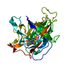
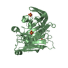
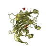
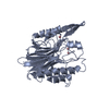
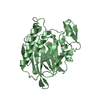
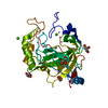
 Bacillus halodurans C-125 (bacteria)
 X-RAY DIFFRACTION /
  Authors
 Citation
 Structure visualization
 Downloads & links
 Other downloads


 PDBj
PDBj

 Assembly


 Mass: 65.409 Da / Num. of mol.: 1 / Source method: obtained synthetically / Formula: Zn
 Mass: 18.015 Da / Num. of mol.: 28 / Source method: isolated from a natural source / Formula: H2O
 Sample preparation
 / Beamline: BL11-1 / Wavelength: 0.92522, 0.97922, 0.97464
 Processing