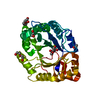
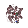
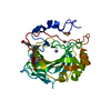
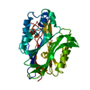
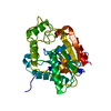
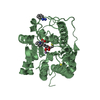
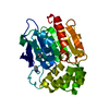
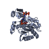
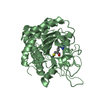
- PDB-4rw3: Structural insights into substrate binding of brown spider venom ... -
+
Open data
ID or keywords:
Loading...
-
Basic information
Entry
Database: PDB / ID: 4rw3
Title
Structural insights into substrate binding of brown spider venom class II phospholipases D
Components
Phospholipase D LiSicTox-alphaIA1bii
Keywords
HYDROLASE / TIM-barrel fold
Function / homology
Function and homology information
phosphoric diester hydrolase activity / lipid catabolic process / toxin activity / Lyases; Phosphorus-oxygen lyases / killing of cells of another organism / lyase activity / extracellular region / metal ion binding Similarity search - Function
Glycerophosphoryl diester phosphodiesterase family / Phosphatidylinositol (PI) phosphodiesterase / PLC-like phosphodiesterase, TIM beta/alpha-barrel domain superfamily / TIM Barrel / Alpha-Beta Barrel / Alpha Beta Similarity search - Domain/homology
PhospholipaseDLiSicTox-alphaIA1bii / PLD / Dermonecrotic toxin / Loxtox i4 / Sphingomyelin phosphodiesterase D 1 / SMD 1 / SMase D 1 / ...PLD / Dermonecrotic toxin / Loxtox i4 / Sphingomyelin phosphodiesterase D 1 / SMD 1 / SMase D 1 / Sphingomyelinase D 1
Mass: 33739.695 Da / Num. of mol.: 1 Source method: isolated from a genetically manipulated source Source: (gene. exp.) Loxosceles intermedia (spider) / Production host: Escherichia coli (E. coli) / References: UniProt: P0CE82, phospholipase D
Resolution: 1.72→30 Å / Cor.coef. Fo:Fc: 0.956 / Cor.coef. Fo:Fc free: 0.936 / SU B: 2.898 / SU ML: 0.091 / Cross valid method: THROUGHOUT / ESU R: 0.134 / ESU R Free: 0.126 / Stereochemistry target values: MAXIMUM LIKELIHOOD / Details: HYDROGENS HAVE BEEN USED IF PRESENT IN THE INPUT
Rfactor
Num. reflection
% reflection
Selection details
Rfree
0.223
2808
10 %
RANDOM
Rwork
0.18295
-
-
-
obs
0.18703
25218
99.83 %
-
Solvent computation
Ion probe radii: 0.8 Å / Shrinkage radii: 0.8 Å / VDW probe radii: 1.2 Å / Solvent model: MASK
 Movie
Movie Controller

 Yorodumi
 Open data
 Basic information
 Components
 Keywords
 Function and homology information
 Loxosceles intermedia (spider)
 X-RAY DIFFRACTION /
  Authors
 Citation
 Structure visualization
 Downloads & links
 Other downloads
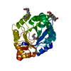


 PDBj
PDBj


 Assembly


 Mass: 24.305 Da / Num. of mol.: 1 / Source method: obtained synthetically / Formula: Mg
 Mass: 106.120 Da / Num. of mol.: 2 / Source method: obtained synthetically / Formula: C4H10O3
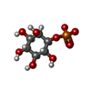
 Mass: 258.120 Da / Num. of mol.: 1 / Source method: obtained synthetically / Formula: C6H11O9P
 Mass: 92.094 Da / Num. of mol.: 1 / Source method: obtained synthetically / Formula: C3H8O3
 Mass: 144.211 Da / Num. of mol.: 1 / Source method: obtained synthetically / Formula: C8H16O2
 Mass: 130.185 Da / Num. of mol.: 1 / Source method: obtained synthetically / Formula: C7H14O2
 Mass: 172.265 Da / Num. of mol.: 2 / Source method: obtained synthetically / Formula: C10H20O2
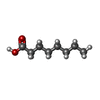
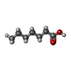
 Mass: 256.424 Da / Num. of mol.: 3 / Source method: obtained synthetically / Formula: C16H32O2
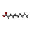
 Mass: 214.344 Da / Num. of mol.: 1 / Source method: obtained synthetically / Formula: C13H26O2
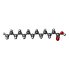
 Sample preparation
 / Beamline: W01B-MX2 / Wavelength: 1.458 Å
 Processing