[English] 日本語
Yorodumi
- PDB-3com: Crystal structure of Mst1 kinase -

+
Open data


ID or keywords:

Loading...

-
Basic information

Entry
Database: PDB / ID: 3com
TitleCrystal structure of Mst1 kinase
ComponentsSerine/threonine-protein kinase 4Serine/threonine-specific protein kinase
Keywords
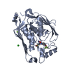
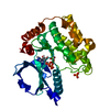
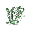
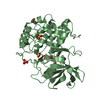
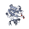
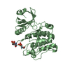
TRANSFERASE / kinase / Mst1 / Serine/threonine-protein kinase 4 / STE20-like kinase / PSI / structural genomics / Protein Structure Initiative / New York SGX Research Center for Structural Genomics / NYSGXRC / Alternative splicing / Apoptosis / ATP-binding / Coiled coil / Cytoplasm / Magnesium / Metal-binding / Nucleotide-binding / Nucleus / Phosphoprotein / Polymorphism
Function / homology
Function and homology information


positive regulation of hepatocyte apoptotic process / cell differentiation involved in embryonic placenta development / regulation of cell differentiation involved in embryonic placenta development / primitive hemopoiesis / neural tube formation / positive regulation of extrinsic apoptotic signaling pathway via death domain receptors / positive regulation of substrate-dependent cell migration, cell attachment to substrate / endocardium development / negative regulation of organ growth / hippo signaling ...positive regulation of hepatocyte apoptotic process / cell differentiation involved in embryonic placenta development / regulation of cell differentiation involved in embryonic placenta development / primitive hemopoiesis / neural tube formation / positive regulation of extrinsic apoptotic signaling pathway via death domain receptors / positive regulation of substrate-dependent cell migration, cell attachment to substrate / endocardium development / negative regulation of organ growth / hippo signaling / Signaling by Hippo / organ growth / branching involved in blood vessel morphogenesis / hepatocyte apoptotic process / regulation of MAPK cascade / extrinsic apoptotic signaling pathway via death domain receptors / canonical Wnt signaling pathway / positive regulation of fat cell differentiation / keratinocyte differentiation / protein serine/threonine kinase activator activity / epithelial cell proliferation / central nervous system development / protein tetramerization / negative regulation of canonical Wnt signaling pathway / cell morphogenesis / protein import into nucleus / negative regulation of epithelial cell proliferation / positive regulation of peptidyl-serine phosphorylation / positive regulation of protein binding / peptidyl-serine phosphorylation / RNA polymerase II-specific DNA-binding transcription factor binding / protein autophosphorylation / protein stabilization / nuclear body / non-specific serine/threonine protein kinase / protein kinase activity / intracellular signal transduction / positive regulation of protein phosphorylation / positive regulation of apoptotic process / protein phosphorylation / protein serine kinase activity / protein serine/threonine kinase activity / apoptotic process / magnesium ion binding / signal transduction / protein homodimerization activity / protein-containing complex / nucleoplasm / ATP binding / identical protein binding / nucleus / cytosol / cytoplasm
Similarity search - Function
Mst1 SARAH domain / C terminal SARAH domain of Mst1 / SARAH domain / SARAH domain profile. / p53-like tetramerisation domain superfamily / Transferase(Phosphotransferase) domain 1 / Transferase(Phosphotransferase); domain 1 / Phosphorylase Kinase; domain 1 / Phosphorylase Kinase; domain 1 / Protein kinase domain ...Mst1 SARAH domain / C terminal SARAH domain of Mst1 / SARAH domain / SARAH domain profile. / p53-like tetramerisation domain superfamily / Transferase(Phosphotransferase) domain 1 / Transferase(Phosphotransferase); domain 1 / Phosphorylase Kinase; domain 1 / Phosphorylase Kinase; domain 1 / Protein kinase domain / Serine/Threonine protein kinases, catalytic domain / Protein kinase, ATP binding site / Protein kinases ATP-binding region signature. / Protein kinase domain profile. / Protein kinase domain / Protein kinase-like domain superfamily / 2-Layer Sandwich / Orthogonal Bundle / Mainly Alpha / Alpha Beta
Similarity search - Domain/homology
Serine/threonine-protein kinase 4
Similarity search - Component
Biological speciesHomo sapiens (human)
MethodX-RAY DIFFRACTION / SYNCHROTRON / MOLECULAR REPLACEMENT / Resolution: 2.2 Å
AuthorsAtwell, S. / Burley, S.K. / Dickey, M. / Leon, B. / Sauder, J.M. / New York SGX Research Center for Structural Genomics (NYSGXRC)
CitationJournal: To be Published
Title: Crystal structure of Mst1 kinase.
Authors: Atwell, S. / Burley, S.K. / Dickey, M. / Leon, B. / Sauder, J.M.
History
DepositionMar 28, 2008Deposition site: RCSB / Processing site: RCSB
Revision 1.0Apr 15, 2008Provider: repository / Type: Initial release
Revision 1.1Jul 13, 2011Group: Version format compliance
Revision 1.2Oct 25, 2017Group: Refinement description / Category: software
Revision 1.3Nov 14, 2018Group: Data collection / Structure summary / Category: audit_author / Item: _audit_author.identifier_ORCID
Revision 1.4Feb 3, 2021Group: Database references / Derived calculations / Structure summary
Category: audit_author / citation_author ...audit_author / citation_author / struct_conn / struct_ref_seq_dif
Item: _audit_author.identifier_ORCID / _citation_author.identifier_ORCID ..._audit_author.identifier_ORCID / _citation_author.identifier_ORCID / _struct_conn.pdbx_leaving_atom_flag / _struct_ref_seq_dif.details
Revision 1.5Aug 30, 2023Group: Data collection / Database references / Refinement description
Category: chem_comp_atom / chem_comp_bond ...chem_comp_atom / chem_comp_bond / database_2 / pdbx_initial_refinement_model
Item: _database_2.pdbx_DOI / _database_2.pdbx_database_accession

-
Structure visualization

Structure viewerMolecule:
MolmilJmol/JSmol

Downloads & links

-
Assembly

Deposited unit
A: Serine/threonine-protein kinase 4
B: Serine/threonine-protein kinase 4


Theoretical massNumber of molelcules
Total (without water)71,5882
Polymers71,5882
Non-polymers00
Water4,161231
1
A: Serine/threonine-protein kinase 4


Theoretical massNumber of molelcules
Total (without water)35,7941
Polymers35,7941
Non-polymers00
Water181
TypeNameSymmetry operationNumber
identity operation1_555x,y,z1
MethodPISA
2
B: Serine/threonine-protein kinase 4


Theoretical massNumber of molelcules
Total (without water)35,7941
Polymers35,7941
Non-polymers00
Water181
TypeNameSymmetry operationNumber
identity operation1_555x,y,z1
MethodPISA
Unit cell
Length a, b, c (Å)74.977, 105.151, 110.955
Angle α, β, γ (deg.)90.00, 90.00, 90.00
Int Tables number19
Space group name H-MP212121

-
Components

#1: Protein Serine/threonine-protein kinase 4 / Serine/threonine-specific protein kinase / STE20-like kinase MST1 / MST-1 / Mammalian STE20-like protein kinase 1 / Serine/threonine-protein kinase Krs-2


Mass: 35793.863 Da / Num. of mol.: 2 / Fragment: Protein kinase domain: Residues 2-311
Source method: isolated from a genetically manipulated source
Source: (gene. exp.) Homo sapiens (human) / Gene: STK4, MST1 / Plasmid: pSGX4 / Species (production host): Escherichia coli / Production host: Escherichia coli BL21(DE3) (bacteria) / Strain (production host): BL21(DE3)
References: UniProt: Q13043, non-specific serine/threonine protein kinase
#2: Water ChemComp-HOH / water / Water


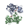
Mass: 18.015 Da / Num. of mol.: 231 / Source method: isolated from a natural source / Formula: H2O

-
Experimental details

-
Experiment

ExperimentMethod: X-RAY DIFFRACTION / Number of used crystals: 1

-
Sample preparation

CrystalDensity Matthews: 3.05 Å3/Da / Density % sol: 59.74 %
Crystal growTemperature: 294 K / Method: vapor diffusion, sitting drop / pH: 10.5
Details: 1700mM Ammonium sulfate, 300mM Lithium sulfate, 100mM CAPS pH 10.5, VAPOR DIFFUSION, SITTING DROP, temperature 294K

-
Data collection

DiffractionMean temperature: 100 K
Diffraction sourceSource: SYNCHROTRON / Site: APS / Beamline: 31-ID / Wavelength: 0.9794 Å
DetectorType: MAR CCD 165 mm / Detector: CCD / Date: Jun 1, 2004 / Details: KV mirrors
RadiationMonochromator: Kohzu 111 diamond / Protocol: SINGLE WAVELENGTH / Monochromatic (M) / Laue (L): M / Scattering type: x-ray
Radiation wavelengthWavelength: 0.9794 Å / Relative weight: 1
ReflectionResolution: 2.2→30 Å / Num. obs: 40513 / % possible obs: 90.2 % / Redundancy: 3.2 % / Biso Wilson estimate: 27.2 Å2 / Rmerge(I) obs: 0.118 / Net I/σ(I): 6.5
Reflection shellResolution: 2.2→2.32 Å / Redundancy: 3.1 % / Rmerge(I) obs: 0.439 / Mean I/σ(I) obs: 2.7 / Num. unique all: 6082 / % possible all: 93.9

-
Processing

Software
NameVersionClassification
MAR345data collection
MOLREPphasing
REFMAC5refinement
MOSFLMdata reduction
SCALAdata scaling
RefinementMethod to determine structure: MOLECULAR REPLACEMENT
Starting model: PDB entry 3CKW
Resolution: 2.2→30 Å / Isotropic thermal model: Isotropic / Stereochemistry target values: Engh & Huber
RfactorNum. reflection% reflectionSelection details
Rfree0.244 2022 -random
Rwork0.208 ---
obs0.208 40443 90.2 %-
Refinement stepCycle: LAST / Resolution: 2.2→30 Å
ProteinNucleic acidLigandSolventTotal
Num. atoms4283 0 0 231 4514
Refine LS restraints
Refine-IDTypeDev ideal
X-RAY DIFFRACTIONr_bond_refined_d0.015
X-RAY DIFFRACTIONr_bond_d_na
X-RAY DIFFRACTIONr_bond_d_prot
X-RAY DIFFRACTIONr_angle_refined_deg1.394
X-RAY DIFFRACTIONr_angle_d_na
X-RAY DIFFRACTIONr_angle_d_prot
X-RAY DIFFRACTIONr_angle_deg
X-RAY DIFFRACTIONr_angle_deg_na
X-RAY DIFFRACTIONr_angle_deg_prot
X-RAY DIFFRACTIONr_dihedral_angle_d
X-RAY DIFFRACTIONr_dihedral_angle_d_na
X-RAY DIFFRACTIONr_dihedral_angle_d_prot
X-RAY DIFFRACTIONr_improper_angle_d
X-RAY DIFFRACTIONr_improper_angle_d_na
X-RAY DIFFRACTIONr_improper_angle_d_prot
X-RAY DIFFRACTIONr_mcbond_it
X-RAY DIFFRACTIONr_mcangle_it
X-RAY DIFFRACTIONr_scbond_it
X-RAY DIFFRACTIONr_scangle_it

+
About Yorodumi

-
News

-
Feb 9, 2022. New format data for meta-information of EMDB entries

New format data for meta-information of EMDB entries

  • Version 3 of the EMDB header file is now the official format.
  • The previous official version 1.9 will be removed from the archive.

Related info.:EMDB header

External links:wwPDB to switch to version 3 of the EMDB data model

-
Aug 12, 2020. Covid-19 info

Covid-19 info

URL: https://pdbj.org/emnavi/covid19.php

New page: Covid-19 featured information page in EM Navigator.

Related info.:Covid-19 info / Mar 5, 2020. Novel coronavirus structure data

+
Mar 5, 2020. Novel coronavirus structure data

Novel coronavirus structure data

Related info.:Yorodumi Speices / Aug 12, 2020. Covid-19 info

External links:COVID-19 featured content - PDBj / Molecule of the Month (242):Coronavirus Proteases

+
Jan 31, 2019. EMDB accession codes are about to change! (news from PDBe EMDB page)

EMDB accession codes are about to change! (news from PDBe EMDB page)

  • The allocation of 4 digits for EMDB accession codes will soon come to an end. Whilst these codes will remain in use, new EMDB accession codes will include an additional digit and will expand incrementally as the available range of codes is exhausted. The current 4-digit format prefixed with “EMD-” (i.e. EMD-XXXX) will advance to a 5-digit format (i.e. EMD-XXXXX), and so on. It is currently estimated that the 4-digit codes will be depleted around Spring 2019, at which point the 5-digit format will come into force.
  • The EM Navigator/Yorodumi systems omit the EMD- prefix.

Related info.:Q: What is EMD? / ID/Accession-code notation in Yorodumi/EM Navigator

External links:EMDB Accession Codes are Changing Soon! / Contact to PDBj

+
Jul 12, 2017. Major update of PDB

Major update of PDB

  • wwPDB released updated PDB data conforming to the new PDBx/mmCIF dictionary.
  • This is a major update changing the version number from 4 to 5, and with Remediation, in which all the entries are updated.
  • In this update, many items about electron microscopy experimental information are reorganized (e.g. em_software).
  • Now, EM Navigator and Yorodumi are based on the updated data.

External links:wwPDB Remediation / Enriched Model Files Conforming to OneDep Data Standards Now Available in the PDB FTP Archive

-
Yorodumi

Thousand views of thousand structures

  • Yorodumi is a browser for structure data from EMDB, PDB, SASBDB, etc.
  • This page is also the successor to EM Navigator detail page, and also detail information page/front-end page for Omokage search.
  • The word "yorodu" (or yorozu) is an old Japanese word meaning "ten thousand". "mi" (miru) is to see.

Related info.:EMDB / PDB / SASBDB / Comparison of 3 databanks / Yorodumi Search / Aug 31, 2016. New EM Navigator & Yorodumi / Yorodumi Papers / Jmol/JSmol / Function and homology information / Changes in new EM Navigator and Yorodumi

Read more