 Movie
Movie Controller

[English] 日本語
 Yorodumi
- PDB-3ch7: Crystal structure of 6-phosphogluconolactonase from Leishmania br... -
+ Open data

- Basic information
| Entry | Database: PDB / ID: 3ch7 | ||||||
|---|---|---|---|---|---|---|---|
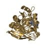
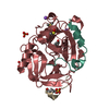
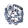
| Title | Crystal structure of 6-phosphogluconolactonase from Leishmania braziliensis | ||||||
 Components Components | 6-phosphogluconolactonase | ||||||
 Keywords Keywords | HYDROLASE / STRUCTURAL GENOMICS / 6-phosphogluconolactonase / leishmaniasis / PSI / Protein Structure Initiative / Structural Genomics of Pathogenic Protozoa Consortium / SGPP | ||||||
| Function / homology |  Function and homology information Function and homology information6-phosphogluconolactonase / 6-phosphogluconolactonase activity / pentose-phosphate shunt / carbohydrate metabolic process Similarity search - Function | ||||||
| Biological species |  Leishmania braziliensis (eukaryote) Leishmania braziliensis (eukaryote) | ||||||
| Method |  X-RAY DIFFRACTION / SYNCHROTRON / MOLECULAR REPLACEMENT / molecular replacement / Resolution: 2.29 Å X-RAY DIFFRACTION / SYNCHROTRON / MOLECULAR REPLACEMENT / molecular replacement / Resolution: 2.29 Å | ||||||
 Authors Authors | Arakaki, T.L. / Merritt, E.A. / Structural Genomics of Pathogenic Protozoa Consortium (SGPP) | ||||||
 Citation Citation | Journal: to be published Title: 6-phosphogluconolactonase from Leishmania braziliensis. Authors: Arakaki, T.L. / Merritt, E.A. | ||||||
| History |
|
- Structure visualization
| Structure viewer | Molecule: MolmilJmol/JSmol |
|---|
- Downloads & links
-Download
| PDBx/mmCIF format | 3ch7.cif.gz | 65.6 KB | Display | PDBx/mmCIF format |
|---|---|---|---|---|
| PDB format | pdb3ch7.ent.gz | 47 KB | Display | PDB format |
| PDBx/mmJSON format | 3ch7.json.gz | Tree view | PDBx/mmJSON format | |
| Others |  Other downloads Other downloads |
-Validation report
| Arichive directory | https://data.pdbj.org/pub/pdb/validation_reports/ch/3ch7ftp://data.pdbj.org/pub/pdb/validation_reports/ch/3ch7 | HTTPS FTP |
|---|
-Related structure data
| Related structure data | 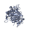 2j0eS S: Starting model for refinement |
|---|---|
| Similar structure data | |
| Other databases |
|


-Links
 PDBj
PDBj- Assembly
| Deposited unit | 
| ||||||||
|---|---|---|---|---|---|---|---|---|---|
| 1 |
| ||||||||
| Unit cell |
|
-Components
| #1: Protein | Mass: 28557.590 Da / Num. of mol.: 1 Source method: isolated from a genetically manipulated source Source: (gene. exp.) Leishmania braziliensis (eukaryote) / Strain: MHOM/BR/75/M2904 / Gene: Lbra003394AAA, LbrM26_V2.2630 / Plasmid: BG1861 / Species (production host): Escherichia coli / Production host:  |
|---|---|
| #2: Water | ChemComp-HOH /  Mass: 18.015 Da / Num. of mol.: 74 / Source method: isolated from a natural source / Formula: H2O Mass: 18.015 Da / Num. of mol.: 74 / Source method: isolated from a natural source / Formula: H2O |
| Sequence details | AUTHORS STATE THAT THE DIFFERENCES BETWEEN THE SEQUENCE OF THEIR PROTEIN AND THE UNIPROT SEQUENCE ...AUTHORS STATE THAT THE DIFFERENCE |
-Experimental details
-Experiment
| Experiment | Method: X-RAY DIFFRACTION / Number of used crystals: 1 |
|---|
- Sample preparation
| Crystal | Density Matthews: 2.72 Å3/Da / Density % sol: 54.85 % |
|---|---|
| Crystal grow | Temperature: 298 K / Method: vapor diffusion / pH: 7.5 Details: 40% PEG 1000, 0.1 M MOPS, 0.1 M NH4H2PO4, pH 7.5, VAPOR DIFFUSION, temperature 298K |
-Data collection
| Diffraction | Mean temperature: 100 K | |||||||||||||||||||||||||||||||||||||||||||||||||||||||||||||||||||||||||||||
|---|---|---|---|---|---|---|---|---|---|---|---|---|---|---|---|---|---|---|---|---|---|---|---|---|---|---|---|---|---|---|---|---|---|---|---|---|---|---|---|---|---|---|---|---|---|---|---|---|---|---|---|---|---|---|---|---|---|---|---|---|---|---|---|---|---|---|---|---|---|---|---|---|---|---|---|---|---|---|
| Diffraction source | Source: SYNCHROTRON / Site: SSRL  / Beamline: BL9-2 / Wavelength: 0.9184 Å / Beamline: BL9-2 / Wavelength: 0.9184 Å | |||||||||||||||||||||||||||||||||||||||||||||||||||||||||||||||||||||||||||||
| Detector | Type: MARMOSAIC 325 mm CCD / Detector: CCD / Date: May 16, 2007 | |||||||||||||||||||||||||||||||||||||||||||||||||||||||||||||||||||||||||||||
| Radiation | Protocol: SINGLE WAVELENGTH / Monochromatic (M) / Laue (L): M / Scattering type: x-ray | |||||||||||||||||||||||||||||||||||||||||||||||||||||||||||||||||||||||||||||
| Radiation wavelength | Wavelength: 0.9184 Å / Relative weight: 1 | |||||||||||||||||||||||||||||||||||||||||||||||||||||||||||||||||||||||||||||
| Reflection | Redundancy: 6.9 % / Av σ(I) over netI: 13.9 / Number: 96585 / Rmerge(I) obs: 0.069 / Χ2: 1.01 / D res high: 2.29 Å / D res low: 50 Å / Num. obs: 13966 / % possible obs: 96.5 | |||||||||||||||||||||||||||||||||||||||||||||||||||||||||||||||||||||||||||||
| Diffraction reflection shell |
| |||||||||||||||||||||||||||||||||||||||||||||||||||||||||||||||||||||||||||||
| Reflection | Resolution: 2.29→50 Å / Num. obs: 13966 / % possible obs: 96.5 % / Redundancy: 6.9 % / Rmerge(I) obs: 0.069 / Χ2: 1.015 / Net I/σ(I): 13.9 | |||||||||||||||||||||||||||||||||||||||||||||||||||||||||||||||||||||||||||||
| Reflection shell |
|
-Phasing
| Phasing | Method: molecular replacement | |||||||||
|---|---|---|---|---|---|---|---|---|---|---|
| Phasing MR |
|
- Processing
| Software |
| |||||||||||||||||||||||||||||||||||||||||||||||||||||||||||||||||||||||||||||||||||||
|---|---|---|---|---|---|---|---|---|---|---|---|---|---|---|---|---|---|---|---|---|---|---|---|---|---|---|---|---|---|---|---|---|---|---|---|---|---|---|---|---|---|---|---|---|---|---|---|---|---|---|---|---|---|---|---|---|---|---|---|---|---|---|---|---|---|---|---|---|---|---|---|---|---|---|---|---|---|---|---|---|---|---|---|---|---|---|
| Refinement | Method to determine structure: MOLECULAR REPLACEMENT Starting model: PDB entry 2J0E Resolution: 2.29→40 Å / Cor.coef. Fo:Fc: 0.958 / Cor.coef. Fo:Fc free: 0.937 / SU B: 6.951 / SU ML: 0.169 / Cross valid method: THROUGHOUT / σ(F): 0 / ESU R: 0.294 / ESU R Free: 0.233 / Stereochemistry target values: MAXIMUM LIKELIHOOD / Details: HYDROGENS HAVE BEEN ADDED IN THE RIDING POSITIONS
| |||||||||||||||||||||||||||||||||||||||||||||||||||||||||||||||||||||||||||||||||||||
| Solvent computation | Ion probe radii: 0.8 Å / Shrinkage radii: 0.8 Å / VDW probe radii: 1.4 Å / Solvent model: MASK | |||||||||||||||||||||||||||||||||||||||||||||||||||||||||||||||||||||||||||||||||||||
| Displacement parameters | Biso mean: 33.788 Å2
| |||||||||||||||||||||||||||||||||||||||||||||||||||||||||||||||||||||||||||||||||||||
| Refinement step | Cycle: LAST / Resolution: 2.29→40 Å
| |||||||||||||||||||||||||||||||||||||||||||||||||||||||||||||||||||||||||||||||||||||
| Refine LS restraints |
| |||||||||||||||||||||||||||||||||||||||||||||||||||||||||||||||||||||||||||||||||||||
| LS refinement shell | Resolution: 2.29→2.345 Å / Total num. of bins used: 20
|
