Mass: 18.015 Da / Num. of mol.: 704 / Source method: isolated from a natural source / Formula: H2O
Has protein modification
Y
Sequence details
THE CONSTRUCT WAS EXPRESSED WITH A PURIFICATION TAG MGSDKIHHHHHHENLYFQG. THE TAG WAS REMOVED WITH ...THE CONSTRUCT WAS EXPRESSED WITH A PURIFICATION TAG MGSDKIHHHHHHENLYFQG. THE TAG WAS REMOVED WITH TEV PROTEASE LEAVING ONLY A GLYCINE (0) FOLLOWED BY THE TARGET SEQUENCE. THE CONSTRUCT CONTAINS RESIDUES 23-537 OF THE TARGET SEQUENCE.
-
Experimental details
-
Experiment
Experiment
Method: X-RAY DIFFRACTION / Number of used crystals: 1
-
Sample preparation
Crystal
Density Matthews: 2.36 Å3/Da / Density % sol: 47.99 %
Crystal grow
Temperature: 277 K / Method: vapor diffusion, sitting drop / pH: 5.6 Details: NANODROP, 0.2M NH4OAc, 30.0% PEG 4000, 0.1M Citrate pH 5.6, VAPOR DIFFUSION, SITTING DROP, temperature 277K
Resolution: 1.7→28.039 Å / Num. obs: 60062 / % possible obs: 98.5 % / Observed criterion σ(I): -3 / Biso Wilson estimate: 15.794 Å2 / Rmerge(I) obs: 0.056 / Net I/σ(I): 10.61
Reflection shell
Resolution (Å)
Rmerge(I) obs
Mean I/σ(I) obs
Num. measured obs
Num. unique obs
Diffraction-ID
% possible all
1.7-1.76
0.328
2.5
21187
11140
1
97.1
1.76-1.83
0.258
3.1
22031
11511
1
99.4
1.83-1.91
0.205
4
21462
11189
1
99.3
1.91-2.02
0.148
5.4
23980
12489
1
99.3
2.02-2.14
0.106
7.3
21043
10937
1
99.4
2.14-2.31
0.081
9.4
22875
11880
1
99.4
2.31-2.54
0.065
11.6
21982
11339
1
99.1
2.54-2.9
0.047
15
22053
11297
1
98.5
2.9-3.66
0.032
20.8
22647
11457
1
97.8
3.66-28.039
0.025
27.4
22465
11225
1
96
-
Phasing
Phasing
Method: MAD
-
Processing
Software
Name
Version
Classification
NB
REFMAC
5.4.0067
refinement
PHENIX
refinement
SHELX
phasing
MolProbity
3beta29
modelbuilding
XSCALE
datascaling
PDB_EXTRACT
3
dataextraction
MAR345
CCD
datacollection
XDS
datareduction
SHELXD
phasing
autoSHARP
phasing
Refinement
Method to determine structure: MAD / Resolution: 1.7→28.039 Å / Cor.coef. Fo:Fc: 0.97 / Cor.coef. Fo:Fc free: 0.96 / SU B: 2.99 / SU ML: 0.052 / TLS residual ADP flag: LIKELY RESIDUAL / Cross valid method: THROUGHOUT / σ(F): 0 / ESU R: 0.086 / ESU R Free: 0.083 Stereochemistry target values: MAXIMUM LIKELIHOOD WITH PHASES Details: 1. HYDROGENS HAVE BEEN ADDED IN THE RIDING POSITIONS. 2. A MET-INHIBITION PROTOCOL WAS USED FOR SELENOMETHIONINE INCORPORATION DURING PROTEIN EXPRESSION. THE OCCUPANCY OF THE SE ATOMS IN THE ...Details: 1. HYDROGENS HAVE BEEN ADDED IN THE RIDING POSITIONS. 2. A MET-INHIBITION PROTOCOL WAS USED FOR SELENOMETHIONINE INCORPORATION DURING PROTEIN EXPRESSION. THE OCCUPANCY OF THE SE ATOMS IN THE MSE RESIDUES WAS REDUCED TO 0.75 FOR THE REDUCED SCATTERING POWER DUE TO PARTIAL S-MET INCORPORATION. 3. ATOM RECORD CONTAINS RESIDUAL B FACTORS ONLY. 4. ACT IS MODELED BASED ON CRYSTALLIZATION CONDITIONS. 5. ZN IS MODELED BASED ON AN X-RAY FLOURESCENCE SCAN, ANOMALOUS DIFFERENCE FOURIERS, AND COORDINATION GEOMETRY. 6. RESIDUE GLY 41 IS A RAMACHANDRAN OUTLIER, AND IS PRESENT IN POOR DENSITY.
Rfactor
Num. reflection
% reflection
Selection details
Rfree
0.166
3037
5.1 %
RANDOM
Rwork
0.14
-
-
-
obs
0.142
60025
99.2 %
-
Solvent computation
Ion probe radii: 0.8 Å / Shrinkage radii: 0.8 Å / VDW probe radii: 1.2 Å / Solvent model: MASK
Displacement parameters
Biso mean: 12.202 Å2
Baniso -1
Baniso -2
Baniso -3
1-
-0.16 Å2
0 Å2
0 Å2
2-
-
0.65 Å2
0 Å2
3-
-
-
-0.49 Å2
Refinement step
Cycle: LAST / Resolution: 1.7→28.039 Å
Protein
Nucleic acid
Ligand
Solvent
Total
Num. atoms
3943
0
5
704
4652
Refine LS restraints
Refine-ID
Type
Dev ideal
Dev ideal target
Number
X-RAY DIFFRACTION
r_bond_refined_d
0.016
0.022
4156
X-RAY DIFFRACTION
r_bond_other_d
0.001
0.02
2719
X-RAY DIFFRACTION
r_angle_refined_deg
1.521
1.933
5694
X-RAY DIFFRACTION
r_angle_other_deg
1.413
3
6655
X-RAY DIFFRACTION
r_dihedral_angle_1_deg
6.169
5
541
X-RAY DIFFRACTION
r_dihedral_angle_2_deg
40.212
25.268
205
X-RAY DIFFRACTION
r_dihedral_angle_3_deg
12.917
15
642
X-RAY DIFFRACTION
r_dihedral_angle_4_deg
18.925
15
17
X-RAY DIFFRACTION
r_chiral_restr
0.1
0.2
611
X-RAY DIFFRACTION
r_gen_planes_refined
0.009
0.021
4806
X-RAY DIFFRACTION
r_gen_planes_other
0.003
0.02
843
X-RAY DIFFRACTION
r_mcbond_it
0.774
1.5
2590
X-RAY DIFFRACTION
r_mcbond_other
0.268
1.5
1048
X-RAY DIFFRACTION
r_mcangle_it
1.289
2
4183
X-RAY DIFFRACTION
r_scbond_it
2.237
3
1566
X-RAY DIFFRACTION
r_scangle_it
3.597
4.5
1496
LS refinement shell
Resolution: 1.7→1.744 Å / Total num. of bins used: 20
Rfactor
Num. reflection
% reflection
Rfree
0.212
228
-
Rwork
0.189
4096
-
all
-
4324
-
obs
-
-
97.61 %
Refinement TLS params.
Method: refined / Origin x: -19.915 Å / Origin y: -52.104 Å / Origin z: 20.37 Å
11
12
13
21
22
23
31
32
33
T
-0.0333 Å2
0.0351 Å2
-0.0053 Å2
-
-0.0276 Å2
-0.0065 Å2
-
-
-0.0482 Å2
L
0.5402 °2
-0.1535 °2
-0.0047 °2
-
0.4763 °2
0.1319 °2
-
-
0.4645 °2
S
0.0633 Å °
0.0265 Å °
0.0087 Å °
-0.0958 Å °
-0.0495 Å °
-0.0043 Å °
-0.0414 Å °
-0.0069 Å °
-0.0138 Å °
+
About Yorodumi
-
News
-
Feb 9, 2022. New format data for meta-information of EMDB entries
New format data for meta-information of EMDB entries
Version 3 of the EMDB header file is now the official format.
The previous official version 1.9 will be removed from the archive.
In the structure databanks used in Yorodumi, some data are registered as the other names, "COVID-19 virus" and "2019-nCoV". Here are the details of the virus and the list of structure data.
Jan 31, 2019. EMDB accession codes are about to change! (news from PDBe EMDB page)
EMDB accession codes are about to change! (news from PDBe EMDB page)
The allocation of 4 digits for EMDB accession codes will soon come to an end. Whilst these codes will remain in use, new EMDB accession codes will include an additional digit and will expand incrementally as the available range of codes is exhausted. The current 4-digit format prefixed with “EMD-” (i.e. EMD-XXXX) will advance to a 5-digit format (i.e. EMD-XXXXX), and so on. It is currently estimated that the 4-digit codes will be depleted around Spring 2019, at which point the 5-digit format will come into force.
The EM Navigator/Yorodumi systems omit the EMD- prefix.
Related info.:Q: What is EMD? / ID/Accession-code notation in Yorodumi/EM Navigator
Yorodumi is a browser for structure data from EMDB, PDB, SASBDB, etc.
This page is also the successor to EM Navigator detail page, and also detail information page/front-end page for Omokage search.
The word "yorodu" (or yorozu) is an old Japanese word meaning "ten thousand". "mi" (miru) is to see.
Related info.:EMDB / PDB / SASBDB / Comparison of 3 databanks / Yorodumi Search / Aug 31, 2016. New EM Navigator & Yorodumi / Yorodumi Papers / Jmol/JSmol / Function and homology information / Changes in new EM Navigator and Yorodumi
 Movie
Movie Controller

 Yorodumi
 Open data
 Basic information
 Components
 Keywords
 Function and homology information
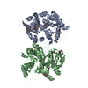
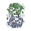
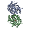
 Bacteroides thetaiotaomicron VPI-5482 (bacteria)
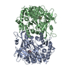
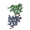
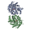
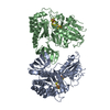
 X-RAY DIFFRACTION /
  Authors
 Citation
 Structure visualization
 Downloads & links
 Other downloads
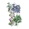

 PDBj
PDBj


 Assembly

 Mass: 65.409 Da / Num. of mol.: 1 / Source method: obtained synthetically / Formula: Zn
 Mass: 59.044 Da / Num. of mol.: 1 / Source method: obtained synthetically / Formula: C2H3O2
 Mass: 18.015 Da / Num. of mol.: 704 / Source method: isolated from a natural source / Formula: H2O
 Sample preparation
 / Beamline: BL9-2 / Wavelength: 0.91837, 0.97911, 0.97922
 Processing