 Movie
Movie Controller

[English] 日本語
 Yorodumi
- PDB-3cg5: Crystal Structure of the Covalent Adduct Formed between TB B-lact... -
+ Open data

- Basic information
| Entry | Database: PDB / ID: 3cg5 | ||||||
|---|---|---|---|---|---|---|---|
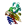
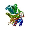
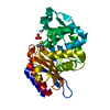
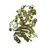
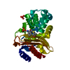
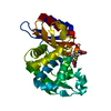
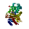
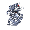
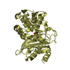
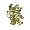
| Title | Crystal Structure of the Covalent Adduct Formed between TB B-lactamase and Clavulanate | ||||||
 Components Components | Beta-lactamase | ||||||
 Keywords Keywords | HYDROLASE / alpha-beta structure / covalent adduct / Antibiotic resistance / Lipoprotein / Membrane / Palmitate | ||||||
| Function / homology |  Function and homology information Function and homology informationbeta-lactam antibiotic catabolic process / beta-lactamase activity / beta-lactamase / periplasmic space / response to antibiotic / extracellular region / plasma membrane Similarity search - Function | ||||||
| Biological species |   Mycobacterium tuberculosis (bacteria) Mycobacterium tuberculosis (bacteria) | ||||||
| Method |  X-RAY DIFFRACTION / SYNCHROTRON / MOLECULAR REPLACEMENT / Resolution: 1.7 Å X-RAY DIFFRACTION / SYNCHROTRON / MOLECULAR REPLACEMENT / Resolution: 1.7 Å | ||||||
 Authors Authors | Tremblay, L.W. / Hugonnet, J.E. / Blanchard, J.S. | ||||||
 Citation Citation | Journal: Biochemistry / Year: 2008 Title: Structure of the covalent adduct formed between Mycobacterium tuberculosis beta-lactamase and clavulanate. Authors: Tremblay, L.W. / Hugonnet, J.E. / Blanchard, J.S. | ||||||
| History |
|
- Structure visualization
| Structure viewer | Molecule: MolmilJmol/JSmol |
|---|
- Downloads & links
-Download
| PDBx/mmCIF format | 3cg5.cif.gz | 70.1 KB | Display | PDBx/mmCIF format |
|---|---|---|---|---|
| PDB format | pdb3cg5.ent.gz | 50.3 KB | Display | PDB format |
| PDBx/mmJSON format | 3cg5.json.gz | Tree view | PDBx/mmJSON format | |
| Others |  Other downloads Other downloads |
-Validation report
| Summary document | 3cg5_validation.pdf.gz | 438.7 KB | Display | wwPDB validaton report |
|---|---|---|---|---|
| Full document | 3cg5_full_validation.pdf.gz | 439.5 KB | Display | |
| Data in XML | 3cg5_validation.xml.gz | 14.4 KB | Display | |
| Data in CIF | 3cg5_validation.cif.gz | 21.7 KB | Display | |
| Arichive directory | https://data.pdbj.org/pub/pdb/validation_reports/cg/3cg5ftp://data.pdbj.org/pub/pdb/validation_reports/cg/3cg5 | HTTPS FTP |
-Related structure data
| Related structure data |  2gdnS S: Starting model for refinement |
|---|---|
| Similar structure data |


-Links
 PDBj
PDBj


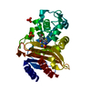
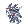
- Assembly
| Deposited unit | 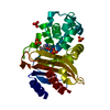
| ||||||||
|---|---|---|---|---|---|---|---|---|---|
| 1 |
| ||||||||
| Unit cell |
|
-Components
| #1: Protein | Mass: 28272.721 Da / Num. of mol.: 1 / Fragment: UNP residues 43-307 Source method: isolated from a genetically manipulated source Source: (gene. exp.) Mycobacterium tuberculosis (bacteria) / Gene: blaA, blaC / Plasmid: pET28a / Production host: References: UniProt: P0C5C1, UniProt: P9WKD3*PLUS, beta-lactamase | ||||||||
|---|---|---|---|---|---|---|---|---|---|
| #2: Chemical | ChemComp-PO4 /   Mass: 94.971 Da / Num. of mol.: 4 / Source method: obtained synthetically / Formula: PO4 Mass: 94.971 Da / Num. of mol.: 4 / Source method: obtained synthetically / Formula: PO4#3: Chemical | ChemComp-ISS / ( | 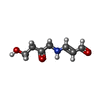  Mass: 157.167 Da / Num. of mol.: 1 / Source method: obtained synthetically / Formula: C7H11NO3 Mass: 157.167 Da / Num. of mol.: 1 / Source method: obtained synthetically / Formula: C7H11NO3#4: Water | ChemComp-HOH / |  Mass: 18.015 Da / Num. of mol.: 271 / Source method: isolated from a natural source / Formula: H2O Mass: 18.015 Da / Num. of mol.: 271 / Source method: isolated from a natural source / Formula: H2OHas protein modification | Y | Nonpolymer details | CLAVULANATE WAS USED IN PROTEIN SOLUTION WHICH UNDERWENT CHEMICAL REACTION AND SUBSEQUENTLY FORMED ...CLAVULANAT | |
-Experimental details
-Experiment
| Experiment | Method: X-RAY DIFFRACTION / Number of used crystals: 1 |
|---|
- Sample preparation
| Crystal | Density Matthews: 2.23 Å3/Da / Density % sol: 44.83 % |
|---|---|
| Crystal grow | Temperature: 298 K / Method: vapor diffusion, hanging drop / pH: 4.1 Details: 0.1 M HEPES pH 7.5, 2 M NH4(H2)PO4, pH 4.1, VAPOR DIFFUSION, HANGING DROP, temperature 298K |
-Data collection
| Diffraction | Mean temperature: 100 K |
|---|---|
| Diffraction source | Source: SYNCHROTRON / Site: NSLS  / Beamline: X12C / Wavelength: 1.1 Å / Beamline: X12C / Wavelength: 1.1 Å |
| Detector | Type: ADSC QUANTUM 210 / Detector: CCD / Date: Aug 8, 2007 |
| Radiation | Monochromator: Graphite / Protocol: SINGLE WAVELENGTH / Monochromatic (M) / Laue (L): M / Scattering type: x-ray |
| Radiation wavelength | Wavelength: 1.1 Å / Relative weight: 1 |
| Reflection | Resolution: 1.7→41.4 Å / Num. all: 27349 / Num. obs: 27349 / % possible obs: 96 % / Observed criterion σ(F): 0 / Observed criterion σ(I): 0 / Redundancy: 3.3 % / Biso Wilson estimate: 15.76 Å2 / Rmerge(I) obs: 0.05 / Net I/σ(I): 18 |
| Reflection shell | Resolution: 1.7→1.74 Å / Redundancy: 3 % / Rmerge(I) obs: 0.4 / Mean I/σ(I) obs: 2 / Num. unique all: 1732 / % possible all: 88 |
- Processing
| Software |
| ||||||||||||||||||||||||||||||||||||||||||||||||||||||||||||||||||||||||||||||||||||||||||
|---|---|---|---|---|---|---|---|---|---|---|---|---|---|---|---|---|---|---|---|---|---|---|---|---|---|---|---|---|---|---|---|---|---|---|---|---|---|---|---|---|---|---|---|---|---|---|---|---|---|---|---|---|---|---|---|---|---|---|---|---|---|---|---|---|---|---|---|---|---|---|---|---|---|---|---|---|---|---|---|---|---|---|---|---|---|---|---|---|---|---|---|
| Refinement | Method to determine structure: MOLECULAR REPLACEMENT Starting model: PDB entry 2GDN Resolution: 1.7→41.4 Å / Cor.coef. Fo:Fc: 0.952 / Cor.coef. Fo:Fc free: 0.929 / SU B: 2.54 / SU ML: 0.081 / Cross valid method: THROUGHOUT / ESU R: 0.116 / ESU R Free: 0.11 / Stereochemistry target values: MAXIMUM LIKELIHOOD
| ||||||||||||||||||||||||||||||||||||||||||||||||||||||||||||||||||||||||||||||||||||||||||
| Solvent computation | Ion probe radii: 0.8 Å / Shrinkage radii: 0.8 Å / VDW probe radii: 1.2 Å / Solvent model: MASK | ||||||||||||||||||||||||||||||||||||||||||||||||||||||||||||||||||||||||||||||||||||||||||
| Displacement parameters | Biso mean: 13.99 Å2
| ||||||||||||||||||||||||||||||||||||||||||||||||||||||||||||||||||||||||||||||||||||||||||
| Refine analyze | Luzzati sigma a obs: 0.1947 Å | ||||||||||||||||||||||||||||||||||||||||||||||||||||||||||||||||||||||||||||||||||||||||||
| Refinement step | Cycle: LAST / Resolution: 1.7→41.4 Å
| ||||||||||||||||||||||||||||||||||||||||||||||||||||||||||||||||||||||||||||||||||||||||||
| Refine LS restraints |
| ||||||||||||||||||||||||||||||||||||||||||||||||||||||||||||||||||||||||||||||||||||||||||
| LS refinement shell | Resolution: 1.7→1.744 Å / Total num. of bins used: 20
|
