 Movie
Movie Controller


+ Open data

- Basic information
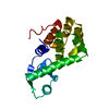
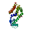
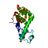
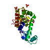
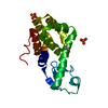
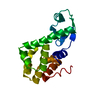
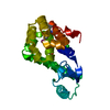
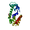
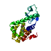
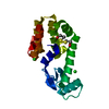
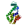
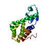
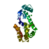
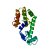
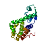
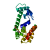
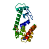
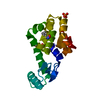
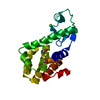
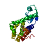
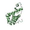
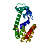
| Entry | Database: PDB / ID: 3cdr | ||||||
|---|---|---|---|---|---|---|---|
| Title | R96Q Mutant of wildtype phage T4 lysozyme at 298 K | ||||||
 Components Components | Lysozyme | ||||||
 Keywords Keywords | HYDROLASE / T4 LYSOZYME / MUTATIONAL ANALYSIS / PROTEIN ENGINEERING / THERMAL STABILITY / PROTEIN STABILITY / PROTEIN ELECTROSTATICS / PROTEIN STRUCTURE / CATION BINDING / CHARGE BURIAL / HYDROGEN BONDING / HELIX DIPOLE / PROTEIN CREVICES / STERIC STRAIN / TEMPERATURE-SENSITIVE MUTANT / Antimicrobial / Bacteriolytic enzyme / Glycosidase | ||||||
| Function / homology |  Function and homology information Function and homology informationviral release from host cell by cytolysis / peptidoglycan catabolic process / cell wall macromolecule catabolic process / lysozyme / lysozyme activity / host cell cytoplasm / defense response to bacterium Similarity search - Function | ||||||
| Biological species |  Bacteriophage T4 (virus) Bacteriophage T4 (virus) | ||||||
| Method |  X-RAY DIFFRACTION / MOLECULAR REPLACEMENT / Resolution: 1.7 Å X-RAY DIFFRACTION / MOLECULAR REPLACEMENT / Resolution: 1.7 Å | ||||||
 Authors Authors | Mooers, B.H.M. | ||||||
 Citation Citation | Journal: Protein Sci. / Year: 2009 Title: Contributions of all 20 amino acids at site 96 to the stability and structure of T4 lysozyme. Authors: Mooers, B.H. / Baase, W.A. / Wray, J.W. / Matthews, B.W. #1: Journal: Protein Sci. / Year: 2009Title: Evaluation at atomic resolution of the role of strain in destabilizing the temperature-sensitive T4 lysozyme mutant Arg 96 --> His. Authors: Mooers, B.H. / Tronrud, D.E. / Matthews, B.W. | ||||||
| History |
|
- Structure visualization
| Structure viewer | Molecule: MolmilJmol/JSmol |
|---|
- Downloads & links
-Download
| PDBx/mmCIF format | 3cdr.cif.gz | 48.1 KB | Display | PDBx/mmCIF format |
|---|---|---|---|---|
| PDB format | pdb3cdr.ent.gz | 33.1 KB | Display | PDB format |
| PDBx/mmJSON format | 3cdr.json.gz | Tree view | PDBx/mmJSON format | |
| Others |  Other downloads Other downloads |
-Validation report
| Arichive directory | https://data.pdbj.org/pub/pdb/validation_reports/cd/3cdrftp://data.pdbj.org/pub/pdb/validation_reports/cd/3cdr | HTTPS FTP |
|---|
-Related structure data
| Related structure data | 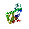 3c7wC 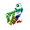 3c7yC  3c7zC 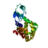 3c80C 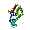 3c81C 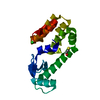 3c82C 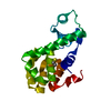 3c83C 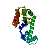 3c8qC 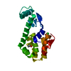 3c8rC  3c8sC 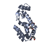 3cdoC 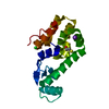 3cdqC  3cdtC 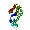 3cdvC 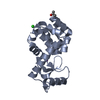 3fi5C 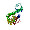 1l63S S: Starting model for refinement C: citing same article ( |
|---|---|
| Similar structure data |


-Links
 PDBj
PDBj


- Assembly
| Deposited unit | 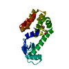
| ||||||||
|---|---|---|---|---|---|---|---|---|---|
| 1 |
| ||||||||
| Unit cell |
|
-Components
| #1: Protein | Mass: 18633.402 Da / Num. of mol.: 1 / Mutation: R96Q Source method: isolated from a genetically manipulated source Source: (gene. exp.) Bacteriophage T4 (virus) / Gene: E / Plasmid: PHS1403 / Production host:  | ||||
|---|---|---|---|---|---|
| #2: Chemical |   Mass: 35.453 Da / Num. of mol.: 2 / Source method: obtained synthetically / Formula: Cl Mass: 35.453 Da / Num. of mol.: 2 / Source method: obtained synthetically / Formula: Cl#3: Chemical | ChemComp-HED / | 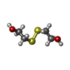  Mass: 154.251 Da / Num. of mol.: 1 / Source method: obtained synthetically / Formula: C4H10O2S2 Mass: 154.251 Da / Num. of mol.: 1 / Source method: obtained synthetically / Formula: C4H10O2S2#4: Water | ChemComp-HOH / |  Mass: 18.015 Da / Num. of mol.: 83 / Source method: isolated from a natural source / Formula: H2O Mass: 18.015 Da / Num. of mol.: 83 / Source method: isolated from a natural source / Formula: H2O |
-Experimental details
-Experiment
| Experiment | Method: X-RAY DIFFRACTION / Number of used crystals: 1 |
|---|
- Sample preparation
| Crystal | Density Matthews: 2.81 Å3/Da / Density % sol: 56.2 % |
|---|---|
| Crystal grow | pH: 6.7 Details: 2 M NA/K PHOSPHATE PH 6.7, 550 MM NACL 50 MM REDUCED BME, 50 MM OXIDIZED BME , VAPOR DIFFUSION, HANGING DROP, TEMPERATURE 277K, pH 6.70 |
-Data collection
| Diffraction | Mean temperature: 298 K |
|---|---|
| Diffraction source | Source: ROTATING ANODE / Type: RIGAKU / Wavelength: 1.5414 |
| Detector | Type: SDMS / Detector: AREA DETECTOR / Date: Sep 4, 1998 / Details: MIRRORS |
| Radiation | Monochromator: GRAPHITE / Protocol: SINGLE WAVELENGTH / Monochromatic (M) / Laue (L): M / Scattering type: x-ray |
| Radiation wavelength | Wavelength: 1.5414 Å / Relative weight: 1 |
| Reflection | Resolution: 1.7→23.215 Å / Num. obs: 21237 / % possible obs: 89 % / Observed criterion σ(I): 2 / Rmerge(I) obs: 0.08 / Rsym value: 0.08 |
| Reflection shell | Resolution: 1.7→1.73 Å / % possible all: 79 |
- Processing
| Software |
| ||||||||||||||||||||||||||||||
|---|---|---|---|---|---|---|---|---|---|---|---|---|---|---|---|---|---|---|---|---|---|---|---|---|---|---|---|---|---|---|---|
| Refinement | Method to determine structure: MOLECULAR REPLACEMENT Starting model: 1L63 Resolution: 1.7→15.46 Å / Cross valid method: F0WT-FOMUT DIFFERENCES MAPS / σ(F): 0 / Stereochemistry target values: TNT5G Details: THR N-CA-C BOND ANGLE WITH A LIBRARY SIGMA OF 2.8 DEGREES. THR158 HAS A DEVIATION OF 2.8 (DEVIATION/SIGMA).
| ||||||||||||||||||||||||||||||
| Solvent computation | Solvent model: BABINET / Bsol: 512.67 Å2 / ksol: 0.9 e/Å3 | ||||||||||||||||||||||||||||||
| Refinement step | Cycle: LAST / Resolution: 1.7→15.46 Å
| ||||||||||||||||||||||||||||||
| Refine LS restraints |
|
