 Movie
Movie Controller


+ Open data

- Basic information
| Entry | Database: PDB / ID: 3c8k | ||||||
|---|---|---|---|---|---|---|---|
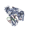
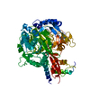
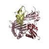
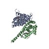
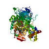
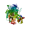
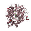
| Title | The crystal structure of Ly49C bound to H-2Kb | ||||||
 Components Components |
| ||||||
 Keywords Keywords | IMMUNE SYSTEM / Natural killer cell receptor / MHC / Virus / Crystal structure / Glycoprotein / Immune response / Membrane / MHC I / Transmembrane / Immunoglobulin domain / Secreted | ||||||
| Function / homology |  Function and homology information Function and homology informationintracellular amino acid homeostasis / RUNX1 regulates transcription of genes involved in differentiation of keratinocytes / phagolysosome / monoatomic ion homeostasis / MHC class Ib protein complex / natural killer cell lectin-like receptor binding / TAP2 binding / TAP1 binding / cis-Golgi network membrane / embryo development ending in birth or egg hatching ...intracellular amino acid homeostasis / RUNX1 regulates transcription of genes involved in differentiation of keratinocytes / phagolysosome / monoatomic ion homeostasis / MHC class Ib protein complex / natural killer cell lectin-like receptor binding / TAP2 binding / TAP1 binding / cis-Golgi network membrane / embryo development ending in birth or egg hatching / Endosomal/Vacuolar pathway / DAP12 interactions / Antigen Presentation: Folding, assembly and peptide loading of class I MHC / ER-Phagosome pathway / response to steroid hormone / DAP12 signaling / Immunoregulatory interactions between a Lymphoid and a non-Lymphoid cell / antigen processing and presentation of exogenous peptide antigen via MHC class Ib / antigen processing and presentation of endogenous peptide antigen via MHC class I via ER pathway, TAP-dependent / TAP complex binding / antigen processing and presentation of exogenous peptide antigen via MHC class I / response to corticosterone / Golgi medial cisterna / inner ear development / regulation of membrane depolarization / CD8 receptor binding / intracellular membrane-bounded organelle / endoplasmic reticulum exit site / TAP binding / MHC class I protein binding / antigen processing and presentation of endogenous peptide antigen via MHC class Ib / antigen processing and presentation of endogenous peptide antigen via MHC class I via ER pathway, TAP-independent / monoatomic ion transport / beta-2-microglobulin binding / cellular defense response / T cell receptor binding / response to progesterone / phagocytic vesicle / Neutrophil degranulation / 14-3-3 protein binding / early endosome lumen / Neutrophil degranulation / lumenal side of endoplasmic reticulum membrane / antigen processing and presentation of exogenous protein antigen via MHC class Ib, TAP-dependent / regulation of iron ion transport / cellular response to iron(III) ion / negative regulation of iron ion transport / negative regulation of forebrain neuron differentiation / response to molecule of bacterial origin / regulation of erythrocyte differentiation / iron ion transport / peptide antigen assembly with MHC class I protein complex / HFE-transferrin receptor complex / MHC class I peptide loading complex / transferrin transport / negative regulation of receptor-mediated endocytosis / cellular response to iron ion / positive regulation of T cell cytokine production / antigen processing and presentation of endogenous peptide antigen via MHC class I / MHC class I protein complex / multicellular organismal-level iron ion homeostasis / peptide antigen assembly with MHC class II protein complex / positive regulation of T cell mediated cytotoxicity / negative regulation of epithelial cell proliferation / cellular response to nicotine / MHC class II protein complex / positive regulation of receptor-mediated endocytosis / negative regulation of neurogenesis / response to estrogen / antigen processing and presentation of exogenous peptide antigen via MHC class II / positive regulation of immune response / peptide antigen binding / phagocytic vesicle membrane / positive regulation of T cell activation / T cell differentiation in thymus / sensory perception of smell / positive regulation of cellular senescence / MHC class II protein complex binding / late endosome membrane / negative regulation of neuron projection development / antimicrobial humoral immune response mediated by antimicrobial peptide / cellular response to lipopolysaccharide / carbohydrate binding / antibacterial humoral response / protein refolding / protein-folding chaperone binding / signaling receptor activity / protease binding / early endosome membrane / amyloid fibril formation / defense response to Gram-negative bacterium / vesicle / protein homotetramerization / early endosome / intracellular iron ion homeostasis / learning or memory / cell adhesion / defense response to bacterium / defense response to Gram-positive bacterium / immune response Similarity search - Function | ||||||
| Biological species |  | ||||||
| Method |  X-RAY DIFFRACTION / SYNCHROTRON / MOLECULAR REPLACEMENT / Resolution: 2.9 Å X-RAY DIFFRACTION / SYNCHROTRON / MOLECULAR REPLACEMENT / Resolution: 2.9 Å | ||||||
 Authors Authors | Deng, L. / Mariuzza, R.A. | ||||||
 Citation Citation | Journal: J.Biol.Chem. / Year: 2008 Title: Molecular architecture of the major histocompatibility complex class I-binding site of Ly49 natural killer cell receptors. Authors: Deng, L. / Cho, S. / Malchiodi, E.L. / Kerzic, M.C. / Dam, J. / Mariuzza, R.A. | ||||||
| History |
|
- Structure visualization
| Structure viewer | Molecule: MolmilJmol/JSmol |
|---|
- Downloads & links
-Download
| PDBx/mmCIF format | 3c8k.cif.gz | 115.9 KB | Display | PDBx/mmCIF format |
|---|---|---|---|---|
| PDB format | pdb3c8k.ent.gz | 90.4 KB | Display | PDB format |
| PDBx/mmJSON format | 3c8k.json.gz | Tree view | PDBx/mmJSON format | |
| Others |  Other downloads Other downloads |
-Validation report
| Arichive directory | https://data.pdbj.org/pub/pdb/validation_reports/c8/3c8kftp://data.pdbj.org/pub/pdb/validation_reports/c8/3c8k | HTTPS FTP |
|---|
-Related structure data
| Related structure data |  3c8jC  3cadC 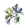 1p4lS C: citing same article ( S: Starting model for refinement |
|---|---|
| Similar structure data |


-Links
 PDBj
PDBj


- Assembly
| Deposited unit | 
| ||||||||
|---|---|---|---|---|---|---|---|---|---|
| 1 |
| ||||||||
| Unit cell |
|
-Components
| #1: Protein | Mass: 31648.322 Da / Num. of mol.: 1 / Fragment: UNP residues 22-295 / Mutation: S171G, E193G, R223K Source method: isolated from a genetically manipulated source Source: (gene. exp.)  |
|---|---|
| #2: Protein | Mass: 11660.350 Da / Num. of mol.: 1 / Fragment: UNP residues 21-119 Source method: isolated from a genetically manipulated source Source: (gene. exp.) |
| #3: Protein/peptide | Mass: 964.137 Da / Num. of mol.: 1 / Source method: obtained synthetically Details: The peptide is chemically synthesized. The sequence occurs naturally in mouse. References: UniProt: P01012*PLUS |
| #4: Protein | Mass: 14915.486 Da / Num. of mol.: 1 / Fragment: UNP residues 142-266 Source method: isolated from a genetically manipulated source Source: (gene. exp.) |
| #5: Water | ChemComp-HOH /  Mass: 18.015 Da / Num. of mol.: 65 / Source method: isolated from a natural source / Formula: H2O Mass: 18.015 Da / Num. of mol.: 65 / Source method: isolated from a natural source / Formula: H2O |
| Has protein modification | Y |
-Experimental details
-Experiment
| Experiment | Method: X-RAY DIFFRACTION / Number of used crystals: 1 |
|---|
- Sample preparation
| Crystal | Density Matthews: 3.16 Å3/Da / Density % sol: 61.02 % |
|---|---|
| Crystal grow | Temperature: 277 K / Method: vapor diffusion / pH: 7.5 Details: 2.0 M ammonium sulfate, 2% (volume/volume) PEG 400 and 0.1 M Hepes, pH 7.5, VAPOR DIFFUSION, temperature 277K |
-Data collection
| Diffraction | Mean temperature: 100 K |
|---|---|
| Diffraction source | Source: SYNCHROTRON / Site: CHESS  / Beamline: F1 / Wavelength: 1 Å / Beamline: F1 / Wavelength: 1 Å |
| Detector | Type: ADSC QUANTUM 4 / Detector: CCD / Date: Feb 1, 2003 |
| Radiation | Protocol: SINGLE WAVELENGTH / Monochromatic (M) / Laue (L): M / Scattering type: x-ray |
| Radiation wavelength | Wavelength: 1 Å / Relative weight: 1 |
| Reflection | Resolution: 2.9→50 Å / Num. obs: 16931 / % possible obs: 97.5 % / Rmerge(I) obs: 0.062 |
| Reflection shell | Resolution: 2.9→2.95 Å / Rmerge(I) obs: 0.325 / % possible all: 97.9 |
- Processing
| Software |
| ||||||||||||||||||||||||||||||||||||||||||||||||||||||||||||||||||||||||||||||||||||||||||||||||||||||||||||||||||||||||||||||||||||||||||||||||||||||||||||||||||||||||||
|---|---|---|---|---|---|---|---|---|---|---|---|---|---|---|---|---|---|---|---|---|---|---|---|---|---|---|---|---|---|---|---|---|---|---|---|---|---|---|---|---|---|---|---|---|---|---|---|---|---|---|---|---|---|---|---|---|---|---|---|---|---|---|---|---|---|---|---|---|---|---|---|---|---|---|---|---|---|---|---|---|---|---|---|---|---|---|---|---|---|---|---|---|---|---|---|---|---|---|---|---|---|---|---|---|---|---|---|---|---|---|---|---|---|---|---|---|---|---|---|---|---|---|---|---|---|---|---|---|---|---|---|---|---|---|---|---|---|---|---|---|---|---|---|---|---|---|---|---|---|---|---|---|---|---|---|---|---|---|---|---|---|---|---|---|---|---|---|---|---|---|---|
| Refinement | Method to determine structure: MOLECULAR REPLACEMENT Starting model: PDB ENTRY 1P4L Resolution: 2.9→30 Å / Cor.coef. Fo:Fc: 0.931 / Cor.coef. Fo:Fc free: 0.882 / SU B: 31.289 / SU ML: 0.294 / Cross valid method: THROUGHOUT / ESU R Free: 0.404 / Stereochemistry target values: MAXIMUM LIKELIHOOD
| ||||||||||||||||||||||||||||||||||||||||||||||||||||||||||||||||||||||||||||||||||||||||||||||||||||||||||||||||||||||||||||||||||||||||||||||||||||||||||||||||||||||||||
| Solvent computation | Ion probe radii: 0.8 Å / Shrinkage radii: 0.8 Å / VDW probe radii: 1.2 Å / Solvent model: MASK | ||||||||||||||||||||||||||||||||||||||||||||||||||||||||||||||||||||||||||||||||||||||||||||||||||||||||||||||||||||||||||||||||||||||||||||||||||||||||||||||||||||||||||
| Displacement parameters | Biso mean: 74.38 Å2
| ||||||||||||||||||||||||||||||||||||||||||||||||||||||||||||||||||||||||||||||||||||||||||||||||||||||||||||||||||||||||||||||||||||||||||||||||||||||||||||||||||||||||||
| Refinement step | Cycle: LAST / Resolution: 2.9→30 Å
| ||||||||||||||||||||||||||||||||||||||||||||||||||||||||||||||||||||||||||||||||||||||||||||||||||||||||||||||||||||||||||||||||||||||||||||||||||||||||||||||||||||||||||
| Refine LS restraints |
| ||||||||||||||||||||||||||||||||||||||||||||||||||||||||||||||||||||||||||||||||||||||||||||||||||||||||||||||||||||||||||||||||||||||||||||||||||||||||||||||||||||||||||
| LS refinement shell | Resolution: 2.9→2.975 Å / Total num. of bins used: 20
|
