 Movie
Movie Controller

[English] 日本語
 Yorodumi
- PDB-3c60: Crystal structure of mouse MHC class II I-Ab/3K peptide complexed... -
+ Open data

- Basic information
| Entry | Database: PDB / ID: 3c60 | ||||||
|---|---|---|---|---|---|---|---|
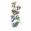
| Title | Crystal structure of mouse MHC class II I-Ab/3K peptide complexed with mouse TCR YAe62 | ||||||
 Components Components |
| ||||||
 Keywords Keywords | SUGAR BINDING PROTEIN/IMMUNE SYSTEM / TCR-pMHC complex / Glycoprotein / Immune response / Membrane / MHC II / Transmembrane / SUGAR BINDING PROTEIN-IMMUNE SYSTEM COMPLEX | ||||||
| Function / homology |  Function and homology information Function and homology informationpositive regulation of antigen processing and presentation / positive regulation of alpha-beta T cell activation / positive regulation of T-helper 1 type immune response / B cell affinity maturation / protein antigen binding / positive regulation of T cell differentiation / response to type II interferon / toxic substance binding / multivesicular body / MHC class II protein complex ...positive regulation of antigen processing and presentation / positive regulation of alpha-beta T cell activation / positive regulation of T-helper 1 type immune response / B cell affinity maturation / protein antigen binding / positive regulation of T cell differentiation / response to type II interferon / toxic substance binding / multivesicular body / MHC class II protein complex / cellular response to type II interferon / antigen processing and presentation of exogenous peptide antigen via MHC class II / peptide antigen binding / adaptive immune response / early endosome / lysosome / external side of plasma membrane / ubiquitin protein ligase binding / cell surface / Golgi apparatus Similarity search - Function | ||||||
| Biological species |  | ||||||
| Method |  X-RAY DIFFRACTION / SYNCHROTRON / MOLECULAR REPLACEMENT / molecular replacement / Resolution: 3.05 Å X-RAY DIFFRACTION / SYNCHROTRON / MOLECULAR REPLACEMENT / molecular replacement / Resolution: 3.05 Å | ||||||
 Authors Authors | Dai, S. | ||||||
 Citation Citation | Journal: Immunity / Year: 2008 Title: Crossreactive T Cells spotlight the germline rules for alphabeta T cell-receptor interactions with MHC molecules. Authors: Dai, S. / Huseby, E.S. / Rubtsova, K. / Scott-Browne, J. / Crawford, F. / Macdonald, W.A. / Marrack, P. / Kappler, J.W. | ||||||
| History |
|
- Structure visualization
| Structure viewer | Molecule: MolmilJmol/JSmol |
|---|
- Downloads & links
-Download
| PDBx/mmCIF format | 3c60.cif.gz | 333.8 KB | Display | PDBx/mmCIF format |
|---|---|---|---|---|
| PDB format | pdb3c60.ent.gz | 268.7 KB | Display | PDB format |
| PDBx/mmJSON format | 3c60.json.gz | Tree view | PDBx/mmJSON format | |
| Others |  Other downloads Other downloads |
-Validation report
| Arichive directory | https://data.pdbj.org/pub/pdb/validation_reports/c6/3c60ftp://data.pdbj.org/pub/pdb/validation_reports/c6/3c60 | HTTPS FTP |
|---|
-Related structure data


-Links
 PDBj
PDBj


- Assembly
| Deposited unit | 
| ||||||||
|---|---|---|---|---|---|---|---|---|---|
| 1 | 
| ||||||||
| 2 | 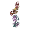
| ||||||||
| Unit cell |
|
-Components
| #1: Protein | Mass: 22109.203 Da / Num. of mol.: 2 Source method: isolated from a genetically manipulated source Source: (gene. exp.) Description: The mouse Va and Vb of YAe62 TCRs fused to human Ca and Cb Plasmid: PET30 / Species (production host): Escherichia coli / Production host:  PDB-3C6O PDB-3C6O#2: Protein | Mass: 26471.277 Da / Num. of mol.: 2 Source method: isolated from a genetically manipulated source Source: (gene. exp.) Description: The mouse Va and Vb of YAe62 TCRs fused to human Ca and Cb Plasmid: PET30 / Species (production host): Escherichia coli / Production host: PDB-3C6O#3: Protein | Mass: 20597.957 Da / Num. of mol.: 2 / Fragment: UNP residues 27-208 Source method: isolated from a genetically manipulated source Source: (gene. exp.)   Spodoptera frugiperda (fall armyworm) / Strain (production host): SF9 / References: UniProt: P14434 Spodoptera frugiperda (fall armyworm) / Strain (production host): SF9 / References: UniProt: P14434#4: Protein | Mass: 24938.830 Da / Num. of mol.: 2 Fragment: Fusion protein of ealpha3K peptide residues 1-13, linker 14-28 and MHC class II Ab UNP residues 30-218 Source method: isolated from a genetically manipulated source Source: (gene. exp.) Spodoptera frugiperda (fall armyworm) / Strain (production host): SF9 / References: UniProt: P14483, PDB-3C6OHas protein modification | Y | Sequence details | BASED ON RELATED ENTRY 1LNU,TWO SEPARATE SEQUENCES, AN EALPHA3KPEPTIDE(1-13) AND LINKER(14-28), ...BASED ON RELATED ENTRY 1LNU,TWO SEPARATE SEQUENCES, AN EALPHA3KPEPTIDE(1-13) AND LINKER(14-28), WHICH START FROM RESIDUE 1 TO RESIDUE 28 WERE ADDED TO THE N-TERMINUS OF THE BETACHAIN OF THE CLASS II MHC IAB OF CHAINS D AND H | |
|---|
-Experimental details
-Experiment
| Experiment | Method: X-RAY DIFFRACTION / Number of used crystals: 1 |
|---|
- Sample preparation
| Crystal | Density Matthews: 3.07 Å3/Da / Density % sol: 59.91 % |
|---|---|
| Crystal grow | Temperature: 298 K / Method: vapor diffusion, hanging drop / pH: 5.2 Details: 15% PEG 4000, pH 5.2, VAPOR DIFFUSION, HANGING DROP, temperature 298K |
-Data collection
| Diffraction | Mean temperature: 100 K |
|---|---|
| Diffraction source | Source: SYNCHROTRON / Site: APS  / Beamline: 19-ID / Wavelength: 0.97918 Å / Beamline: 19-ID / Wavelength: 0.97918 Å |
| Detector | Type: ADSC QUANTUM 315 / Detector: CCD / Date: Apr 9, 2007 / Details: Mirrors |
| Radiation | Monochromator: Si 111 / Protocol: SINGLE WAVELENGTH / Monochromatic (M) / Laue (L): M / Scattering type: x-ray |
| Radiation wavelength | Wavelength: 0.97918 Å / Relative weight: 1 |
| Reflection | Resolution: 3.05→50 Å / Num. obs: 43981 / % possible obs: 97.6 % / Observed criterion σ(I): -3 / Redundancy: 4.5 % / Biso Wilson estimate: 85.8 Å2 / Rmerge(I) obs: 0.087 / Χ2: 1.918 / Net I/σ(I): 13 |
| Reflection shell | Resolution: 3.05→3.16 Å / Redundancy: 4.3 % / Rmerge(I) obs: 0.692 / Mean I/σ(I) obs: 2.9 / Num. unique all: 4349 / Χ2: 1.831 / % possible all: 97.7 |
-Phasing
| Phasing | Method: molecular replacement |
|---|
- Processing
| Software |
| ||||||||||||||||||||||||||||||||||||||||
|---|---|---|---|---|---|---|---|---|---|---|---|---|---|---|---|---|---|---|---|---|---|---|---|---|---|---|---|---|---|---|---|---|---|---|---|---|---|---|---|---|---|
| Refinement | Method to determine structure: MOLECULAR REPLACEMENT / Resolution: 3.05→40.92 Å / Rfactor Rfree error: 0.007 / Data cutoff high absF: 2141643.25 / Data cutoff low absF: 0 / Isotropic thermal model: RESTRAINED / Cross valid method: THROUGHOUT / σ(F): 0
| ||||||||||||||||||||||||||||||||||||||||
| Solvent computation | Solvent model: FLAT MODEL / Bsol: 39.29 Å2 / ksol: 0.299 e/Å3 | ||||||||||||||||||||||||||||||||||||||||
| Displacement parameters | Biso mean: 81.2 Å2
| ||||||||||||||||||||||||||||||||||||||||
| Refine analyze |
| ||||||||||||||||||||||||||||||||||||||||
| Refinement step | Cycle: LAST / Resolution: 3.05→40.92 Å
| ||||||||||||||||||||||||||||||||||||||||
| Refine LS restraints |
| ||||||||||||||||||||||||||||||||||||||||
| LS refinement shell | Resolution: 3.05→3.24 Å / Rfactor Rfree error: 0.025 / Total num. of bins used: 6
| ||||||||||||||||||||||||||||||||||||||||
| Xplor file |
|
