 Movie
Movie Controller

[English] 日本語
 Yorodumi
- PDB-3bzi: Molecular and structural basis of polo-like kinase 1 substrate re... -
+ Open data

- Basic information
| Entry | Database: PDB / ID: 3bzi | |||||||||
|---|---|---|---|---|---|---|---|---|---|---|
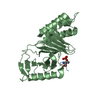
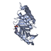
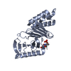
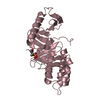
| Title | Molecular and structural basis of polo-like kinase 1 substrate recognition: Implications in centrosomal localization | |||||||||
 Components Components |
| |||||||||
 Keywords Keywords | TRANSFERASE / Kinase / cell cycle / localization / ATP-binding / Nucleotide-binding / Nucleus / Phosphoprotein / Serine/threonine-protein kinase | |||||||||
| Function / homology |  Function and homology information Function and homology informationpositive regulation of G2/MI transition of meiotic cell cycle / positive regulation of mitotic nuclear envelope disassembly / Mitotic Telophase/Cytokinesis / regulation of protein localization to cell cortex / Mitotic Metaphase/Anaphase Transition / synaptonemal complex disassembly / Activation of NIMA Kinases NEK9, NEK6, NEK7 / polo kinase / Phosphorylation of Emi1 / homologous chromosome segregation ...positive regulation of G2/MI transition of meiotic cell cycle / positive regulation of mitotic nuclear envelope disassembly / Mitotic Telophase/Cytokinesis / regulation of protein localization to cell cortex / Mitotic Metaphase/Anaphase Transition / synaptonemal complex disassembly / Activation of NIMA Kinases NEK9, NEK6, NEK7 / polo kinase / Phosphorylation of Emi1 / homologous chromosome segregation / mitotic nuclear membrane disassembly / metaphase/anaphase transition of mitotic cell cycle / female meiosis chromosome segregation / anaphase-promoting complex binding / synaptonemal complex / Phosphorylation of the APC/C / astral microtubule organization / Golgi inheritance / mitotic cleavage furrow formation / outer kinetochore / microtubule bundle formation / mitotic chromosome condensation / double-strand break repair via alternative nonhomologous end joining / regulation of cyclin-dependent protein serine/threonine kinase activity / regulation of mitotic spindle assembly / Polo-like kinase mediated events / Golgi Cisternae Pericentriolar Stack Reorganization / positive regulation of mitotic metaphase/anaphase transition / TP53 regulates transcription of additional cell cycle genes whose exact role in the p53 pathway remain uncertain / positive regulation of ubiquitin-dependent protein catabolic process / centrosome cycle / regulation of mitotic metaphase/anaphase transition / sister chromatid cohesion / regulation of mitotic nuclear division / WW domain binding / Deregulated CDK5 triggers multiple neurodegenerative pathways in Alzheimer's disease models / mitotic spindle pole / mitotic spindle assembly checkpoint signaling / regulation of mitotic cell cycle phase transition / spindle midzone / regulation of anaphase-promoting complex-dependent catabolic process / mitotic G2 DNA damage checkpoint signaling / mitotic cytokinesis / mitotic sister chromatid segregation / positive regulation of G2/M transition of mitotic cell cycle / negative regulation of double-strand break repair via homologous recombination / Activation of ATR in response to replication stress / Regulation of MITF-M-dependent genes involved in cell cycle and proliferation / Cyclin A/B1/B2 associated events during G2/M transition / Chk1/Chk2(Cds1) mediated inactivation of Cyclin B:Cdk1 complex / RHO GTPases activate PKNs / protein localization to chromatin / phosphoprotein phosphatase activity / protein-tyrosine-phosphatase / Loss of Nlp from mitotic centrosomes / Loss of proteins required for interphase microtubule organization from the centrosome / Amplification of signal from unattached kinetochores via a MAD2 inhibitory signal / Recruitment of mitotic centrosome proteins and complexes / protein tyrosine phosphatase activity / Recruitment of NuMA to mitotic centrosomes / Anchoring of the basal body to the plasma membrane / TP53 Regulates Transcription of Genes Involved in G2 Cell Cycle Arrest / Mitotic Prometaphase / regulation of mitotic cell cycle / EML4 and NUDC in mitotic spindle formation / AURKA Activation by TPX2 / Resolution of Sister Chromatid Cohesion / mitotic spindle organization / Condensation of Prophase Chromosomes / regulation of cytokinesis / protein serine/threonine kinase binding / establishment of protein localization / centriole / RHO GTPases Activate Formins / mitochondrial intermembrane space / APC/C:Cdh1 mediated degradation of Cdc20 and other APC/C:Cdh1 targeted proteins in late mitosis/early G1 / positive regulation of protein localization to nucleus / kinetochore / G2/M transition of mitotic cell cycle / spindle / centriolar satellite / spindle pole / The role of GTSE1 in G2/M progression after G2 checkpoint / Separation of Sister Chromatids / Regulation of PLK1 Activity at G2/M Transition / double-strand break repair / mitotic cell cycle / positive regulation of proteasomal ubiquitin-dependent protein catabolic process / microtubule cytoskeleton / midbody / microtubule binding / protein phosphorylation / protein kinase activity / cell population proliferation / regulation of cell cycle / nuclear speck / protein serine kinase activity / cell division / protein serine/threonine kinase activity / centrosome Similarity search - Function | |||||||||
| Biological species |  Homo sapiens (human) Homo sapiens (human) | |||||||||
| Method |  X-RAY DIFFRACTION / SYNCHROTRON / MOLECULAR REPLACEMENT / Resolution: 2.1 Å X-RAY DIFFRACTION / SYNCHROTRON / MOLECULAR REPLACEMENT / Resolution: 2.1 Å | |||||||||
 Authors Authors | Garcia-Alvarez, B. / de Carcer, G. / Ibanez, S. / Bragado-Nilsson, E. / Montoya, G. | |||||||||
 Citation Citation | Journal: Proc.Natl.Acad.Sci.Usa / Year: 2007 Title: Molecular and structural basis of polo-like kinase 1 substrate recognition: Implications in centrosomal localization. Authors: Garcia-Alvarez, B. / de Carcer, G. / Ibanez, S. / Bragado-Nilsson, E. / Montoya, G. | |||||||||
| History |
|
- Structure visualization
| Structure viewer | Molecule: MolmilJmol/JSmol |
|---|
- Downloads & links
-Download
| PDBx/mmCIF format | 3bzi.cif.gz | 65 KB | Display | PDBx/mmCIF format |
|---|---|---|---|---|
| PDB format | pdb3bzi.ent.gz | 46.4 KB | Display | PDB format |
| PDBx/mmJSON format | 3bzi.json.gz | Tree view | PDBx/mmJSON format | |
| Others |  Other downloads Other downloads |
-Validation report
| Arichive directory | https://data.pdbj.org/pub/pdb/validation_reports/bz/3bziftp://data.pdbj.org/pub/pdb/validation_reports/bz/3bzi | HTTPS FTP |
|---|
-Related structure data
| Related structure data |  2ogqC  2ojxC  1owlS S: Starting model for refinement C: citing same article ( |
|---|---|
| Similar structure data |


-Links
 PDBj
PDBj


- Assembly
| Deposited unit | 
| ||||||||
|---|---|---|---|---|---|---|---|---|---|
| 1 |
| ||||||||
| Unit cell |
|
-Components
| #1: Protein | Mass: 27502.334 Da / Num. of mol.: 1 / Fragment: Polo Box Domain Source method: isolated from a genetically manipulated source Source: (gene. exp.) Homo sapiens (human) / Gene: PLK1 / Plasmid: pGEX-6-1P / Production host:  |
|---|---|
| #2: Protein/peptide | Mass: 997.062 Da / Num. of mol.: 1 / Source method: obtained synthetically / Details: synthetic peptide / References: UniProt: P30307 |
| #3: Chemical | ChemComp-FMT /   Mass: 46.025 Da / Num. of mol.: 1 / Source method: obtained synthetically / Formula: CH2O2 Mass: 46.025 Da / Num. of mol.: 1 / Source method: obtained synthetically / Formula: CH2O2 |
| #4: Water | ChemComp-HOH /  Mass: 18.015 Da / Num. of mol.: 172 / Source method: isolated from a natural source / Formula: H2O Mass: 18.015 Da / Num. of mol.: 172 / Source method: isolated from a natural source / Formula: H2O |
| Has protein modification | Y |
-Experimental details
-Experiment
| Experiment | Method: X-RAY DIFFRACTION / Number of used crystals: 1 |
|---|
- Sample preparation
| Crystal | Density Matthews: 2.01 Å3/Da / Density % sol: 38.95 % |
|---|---|
| Crystal grow | Temperature: 298 K / Method: vapor diffusion / pH: 7.5 Details: 100mM HEPES pH 7.5, 2M ammonium formate, VAPOR DIFFUSION, temperature 298K |
-Data collection
| Diffraction | Mean temperature: 100 K |
|---|---|
| Diffraction source | Source: SYNCHROTRON / Site: ESRF  / Beamline: ID14-4 / Wavelength: 0.979 Å / Beamline: ID14-4 / Wavelength: 0.979 Å |
| Detector | Type: ADSC QUANTUM 4 / Detector: CCD |
| Radiation | Protocol: SINGLE WAVELENGTH / Monochromatic (M) / Laue (L): M / Scattering type: x-ray |
| Radiation wavelength | Wavelength: 0.979 Å / Relative weight: 1 |
| Reflection | Resolution: 2.1→36.84 Å / Num. all: 13378 / Num. obs: 12144 / % possible obs: 90.77 % |
- Processing
| Software |
| ||||||||||||||||||||||||||||||||||||||||||||||||||||||||||||||||||||||||||||||||||||||||||
|---|---|---|---|---|---|---|---|---|---|---|---|---|---|---|---|---|---|---|---|---|---|---|---|---|---|---|---|---|---|---|---|---|---|---|---|---|---|---|---|---|---|---|---|---|---|---|---|---|---|---|---|---|---|---|---|---|---|---|---|---|---|---|---|---|---|---|---|---|---|---|---|---|---|---|---|---|---|---|---|---|---|---|---|---|---|---|---|---|---|---|---|
| Refinement | Method to determine structure: MOLECULAR REPLACEMENT Starting model: 1owl Resolution: 2.1→36.84 Å / Cor.coef. Fo:Fc: 0.951 / Cor.coef. Fo:Fc free: 0.907 / SU B: 5.194 / SU ML: 0.138 / Cross valid method: THROUGHOUT / ESU R: 0.275 / ESU R Free: 0.222 / Stereochemistry target values: MAXIMUM LIKELIHOOD
| ||||||||||||||||||||||||||||||||||||||||||||||||||||||||||||||||||||||||||||||||||||||||||
| Solvent computation | Ion probe radii: 0.8 Å / Shrinkage radii: 0.8 Å / VDW probe radii: 1.2 Å / Solvent model: BABINET MODEL WITH MASK | ||||||||||||||||||||||||||||||||||||||||||||||||||||||||||||||||||||||||||||||||||||||||||
| Displacement parameters | Biso mean: 20.739 Å2
| ||||||||||||||||||||||||||||||||||||||||||||||||||||||||||||||||||||||||||||||||||||||||||
| Refinement step | Cycle: LAST / Resolution: 2.1→36.84 Å
| ||||||||||||||||||||||||||||||||||||||||||||||||||||||||||||||||||||||||||||||||||||||||||
| Refine LS restraints |
| ||||||||||||||||||||||||||||||||||||||||||||||||||||||||||||||||||||||||||||||||||||||||||
| LS refinement shell | Resolution: 2.097→2.152 Å / Total num. of bins used: 20
|
