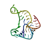
- PDB-3bs5: Crystal Structure of hCNK2-SAM/dHYP-SAM Complex -
+
Open data
ID or keywords:
Loading...
-
Basic information
Entry
Database: PDB / ID: 3bs5
Title
Crystal Structure of hCNK2-SAM/dHYP-SAM Complex
Components
Connector enhancer of kinase suppressor of ras 2
Protein aveugle
Keywords
Signaling PROTEIN/membrane protein / Sterile alpha motif / SAM domain / SAM domain dimer / SAM domain complex / Cytoplasm / Membrane / Sensory transduction / Vision / Alternative splicing / Coiled coil / Phosphoprotein / Polymorphism / Signaling PROTEIN-membrane protein COMPLEX
Function / homology
Function and homology information
Phosphorylation of CI / compound eye cone cell differentiation / postsynaptic specialization organization / Phosphorylation of SMO / Phosphorylation of PER and TIM / compound eye photoreceptor cell differentiation / eye photoreceptor cell differentiation / extrinsic component of postsynaptic density membrane / positive regulation of Ras protein signal transduction / enzyme-linked receptor protein signaling pathway ...Phosphorylation of CI / compound eye cone cell differentiation / postsynaptic specialization organization / Phosphorylation of SMO / Phosphorylation of PER and TIM / compound eye photoreceptor cell differentiation / eye photoreceptor cell differentiation / extrinsic component of postsynaptic density membrane / positive regulation of Ras protein signal transduction / enzyme-linked receptor protein signaling pathway / regulation of signal transduction / visual perception / cell surface receptor protein tyrosine kinase signaling pathway / Signaling by high-kinase activity BRAF mutants / MAP2K and MAPK activation / epidermal growth factor receptor signaling pathway / cytoplasmic side of plasma membrane / Signaling by RAF1 mutants / Signaling by moderate kinase activity BRAF mutants / Paradoxical activation of RAF signaling by kinase inactive BRAF / Signaling downstream of RAS mutants / Signaling by BRAF and RAF1 fusions / scaffold protein binding / protein-macromolecule adaptor activity / positive regulation of ERK1 and ERK2 cascade / intracellular signal transduction / neuronal cell body / protein kinase binding / glutamatergic synapse / extracellular exosome / plasma membrane / cytoplasm / cytosol Similarity search - Function
Connector enhancer of kinase suppressor of ras 2/3 domain / Connector enhancer of kinase suppressor of ras 2/3 domain / Aveugle-like, SAM domain / : / CRIC domain / : / Connector enhancer of kinase suppressor of ras / CRIC domain profile. / : / Transcription Factor, Ets-1 ...Connector enhancer of kinase suppressor of ras 2/3 domain / Connector enhancer of kinase suppressor of ras 2/3 domain / Aveugle-like, SAM domain / : / CRIC domain / : / Connector enhancer of kinase suppressor of ras / CRIC domain profile. / : / Transcription Factor, Ets-1 / SAM domain (Sterile alpha motif) / SAM domain (Sterile alpha motif) / SAM domain profile. / Sterile alpha motif. / PH domain / Sterile alpha motif domain / Sterile alpha motif/pointed domain superfamily / PH domain profile. / PDZ domain / Pleckstrin homology domain. / Pleckstrin homology domain / PDZ domain profile. / Domain present in PSD-95, Dlg, and ZO-1/2. / PDZ domain / PDZ superfamily / DNA polymerase; domain 1 / PH-like domain superfamily / Orthogonal Bundle / Mainly Alpha Similarity search - Domain/homology
Monochromator: Two independent arrays of pairs of vertical and horizontal slit blade pairs Protocol: SINGLE WAVELENGTH / Monochromatic (M) / Laue (L): M / Scattering type: x-ray
Method to determine structure: SAD / Resolution: 2→19.87 Å / Cor.coef. Fo:Fc: 0.941 / Cor.coef. Fo:Fc free: 0.925 / SU B: 3.228 / SU ML: 0.093 / Cross valid method: THROUGHOUT / σ(F): 0 / ESU R: 0.167 / ESU R Free: 0.149 Stereochemistry target values: MAXIMUM LIKELIHOOD WITH PHASES Details: HYDROGENS HAVE BEEN ADDED IN THE RIDING POSITIONS
Rfactor
Num. reflection
% reflection
Selection details
Rfree
0.24
786
4.9 %
RANDOM
Rwork
0.212
-
-
-
all
0.214
-
-
-
obs
0.214
15619
99.67 %
-
Solvent computation
Ion probe radii: 0.8 Å / Shrinkage radii: 0.8 Å / VDW probe radii: 1.4 Å / Solvent model: MASK
Displacement parameters
Biso mean: 32.47 Å2
Baniso -1
Baniso -2
Baniso -3
1-
-0.05 Å2
0 Å2
0 Å2
2-
-
0.04 Å2
0 Å2
3-
-
-
0.01 Å2
Refinement step
Cycle: LAST / Resolution: 2→19.87 Å
Protein
Nucleic acid
Ligand
Solvent
Total
Num. atoms
1299
0
0
82
1381
Refine LS restraints
Refine-ID
Type
Dev ideal
Dev ideal target
Number
X-RAY DIFFRACTION
r_bond_refined_d
0.021
0.021
1304
X-RAY DIFFRACTION
r_angle_refined_deg
1.555
1.946
1756
X-RAY DIFFRACTION
r_dihedral_angle_1_deg
4.884
5
152
X-RAY DIFFRACTION
r_dihedral_angle_2_deg
36.488
23.571
70
X-RAY DIFFRACTION
r_dihedral_angle_3_deg
14.331
15
251
X-RAY DIFFRACTION
r_dihedral_angle_4_deg
14.88
15
14
X-RAY DIFFRACTION
r_chiral_restr
0.113
0.2
192
X-RAY DIFFRACTION
r_gen_planes_refined
0.008
0.02
974
X-RAY DIFFRACTION
r_nbd_refined
0.24
0.2
586
X-RAY DIFFRACTION
r_nbtor_refined
0.305
0.2
903
X-RAY DIFFRACTION
r_xyhbond_nbd_refined
0.249
0.2
101
X-RAY DIFFRACTION
r_symmetry_vdw_refined
0.203
0.2
21
X-RAY DIFFRACTION
r_symmetry_hbond_refined
0.149
0.2
12
X-RAY DIFFRACTION
r_mcbond_it
1.566
1.5
796
X-RAY DIFFRACTION
r_mcangle_it
2.273
2
1227
X-RAY DIFFRACTION
r_scbond_it
3.644
3
599
X-RAY DIFFRACTION
r_scangle_it
5.745
4.5
529
LS refinement shell
Resolution: 2→2.055 Å / Total num. of bins used: 20
Rfactor
Num. reflection
% reflection
Rfree
0.245
56
-
Rwork
0.227
1036
-
all
-
1092
-
obs
-
-
97.59 %
+
About Yorodumi
-
News
-
Feb 9, 2022. New format data for meta-information of EMDB entries
New format data for meta-information of EMDB entries
Version 3 of the EMDB header file is now the official format.
The previous official version 1.9 will be removed from the archive.
In the structure databanks used in Yorodumi, some data are registered as the other names, "COVID-19 virus" and "2019-nCoV". Here are the details of the virus and the list of structure data.
Jan 31, 2019. EMDB accession codes are about to change! (news from PDBe EMDB page)
EMDB accession codes are about to change! (news from PDBe EMDB page)
The allocation of 4 digits for EMDB accession codes will soon come to an end. Whilst these codes will remain in use, new EMDB accession codes will include an additional digit and will expand incrementally as the available range of codes is exhausted. The current 4-digit format prefixed with “EMD-” (i.e. EMD-XXXX) will advance to a 5-digit format (i.e. EMD-XXXXX), and so on. It is currently estimated that the 4-digit codes will be depleted around Spring 2019, at which point the 5-digit format will come into force.
The EM Navigator/Yorodumi systems omit the EMD- prefix.
Related info.:Q: What is EMD? / ID/Accession-code notation in Yorodumi/EM Navigator
Yorodumi is a browser for structure data from EMDB, PDB, SASBDB, etc.
This page is also the successor to EM Navigator detail page, and also detail information page/front-end page for Omokage search.
The word "yorodu" (or yorozu) is an old Japanese word meaning "ten thousand". "mi" (miru) is to see.
Related info.:EMDB / PDB / SASBDB / Comparison of 3 databanks / Yorodumi Search / Aug 31, 2016. New EM Navigator & Yorodumi / Yorodumi Papers / Jmol/JSmol / Function and homology information / Changes in new EM Navigator and Yorodumi
 Movie
Movie Controller


 Open data
 Basic information
 Components
 Keywords
 Function and homology information
 Homo sapiens (human)
 X-RAY DIFFRACTION /
  Authors
 Citation
 Structure visualization
 Downloads & links
 Other downloads


 PDBj
PDBj
 Assembly
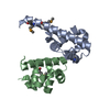

 Mass: 18.015 Da / Num. of mol.: 82 / Source method: isolated from a natural source / Formula: H2O
 Sample preparation
 / Beamline: 24-ID-C / Wavelength: 0.9792 Å
 Processing