Phosphorylation of CI / compound eye cone cell differentiation / Phosphorylation of SMO / Phosphorylation of PER and TIM / compound eye photoreceptor cell differentiation / eye photoreceptor cell differentiation / positive regulation of Ras protein signal transduction / visual perception / cell surface receptor protein tyrosine kinase signaling pathway / epidermal growth factor receptor signaling pathway ...Phosphorylation of CI / compound eye cone cell differentiation / Phosphorylation of SMO / Phosphorylation of PER and TIM / compound eye photoreceptor cell differentiation / eye photoreceptor cell differentiation / positive regulation of Ras protein signal transduction / visual perception / cell surface receptor protein tyrosine kinase signaling pathway / epidermal growth factor receptor signaling pathway / cytoplasmic side of plasma membrane / scaffold protein binding / positive regulation of ERK1 and ERK2 cascade / cytoplasm / cytosol Similarity search - Function
Method to determine structure: MOLECULAR REPLACEMENT / Resolution: 1.9→19.88 Å / Cor.coef. Fo:Fc: 0.924 / Cor.coef. Fo:Fc free: 0.9 / SU B: 3.851 / SU ML: 0.116 / Cross valid method: THROUGHOUT / σ(F): 0 / ESU R: 0.189 / ESU R Free: 0.173 Stereochemistry target values: MAXIMUM LIKELIHOOD WITH PHASES Details: HYDROGENS HAVE BEEN ADDED IN THE RIDING POSITIONS
Rfactor
Num. reflection
% reflection
Selection details
Rfree
0.265
735
5 %
RANDOM
Rwork
0.214
-
-
-
all
0.217
14633
-
-
obs
0.217
14633
99.6 %
-
Solvent computation
Ion probe radii: 0.8 Å / Shrinkage radii: 0.8 Å / VDW probe radii: 1.4 Å / Solvent model: MASK
Displacement parameters
Biso mean: 22.095 Å2
Baniso -1
Baniso -2
Baniso -3
1-
0.01 Å2
0 Å2
0 Å2
2-
-
-0.01 Å2
0 Å2
3-
-
-
0 Å2
Refinement step
Cycle: LAST / Resolution: 1.9→19.88 Å
Protein
Nucleic acid
Ligand
Solvent
Total
Num. atoms
1276
0
0
290
1566
Refine LS restraints
Refine-ID
Type
Dev ideal
Dev ideal target
Number
X-RAY DIFFRACTION
r_bond_refined_d
0.018
0.021
1309
X-RAY DIFFRACTION
r_angle_refined_deg
1.408
1.926
1764
X-RAY DIFFRACTION
r_dihedral_angle_1_deg
4.839
5
149
X-RAY DIFFRACTION
r_dihedral_angle_2_deg
33.277
22.113
71
X-RAY DIFFRACTION
r_dihedral_angle_3_deg
15.96
15
248
X-RAY DIFFRACTION
r_dihedral_angle_4_deg
23.04
15
18
X-RAY DIFFRACTION
r_chiral_restr
0.101
0.2
191
X-RAY DIFFRACTION
r_gen_planes_refined
0.007
0.02
980
X-RAY DIFFRACTION
r_nbd_refined
0.326
0.2
772
X-RAY DIFFRACTION
r_nbtor_refined
0.313
0.2
908
X-RAY DIFFRACTION
r_xyhbond_nbd_refined
0.315
0.2
235
X-RAY DIFFRACTION
r_symmetry_vdw_refined
0.412
0.2
51
X-RAY DIFFRACTION
r_symmetry_hbond_refined
0.285
0.2
33
X-RAY DIFFRACTION
r_mcbond_it
1.272
1.5
774
X-RAY DIFFRACTION
r_mcangle_it
1.942
2
1208
X-RAY DIFFRACTION
r_scbond_it
3.399
3
637
X-RAY DIFFRACTION
r_scangle_it
5.043
4.5
556
LS refinement shell
Resolution: 1.9→1.949 Å / Total num. of bins used: 20
Rfactor
Num. reflection
% reflection
Rfree
0.306
52
-
Rwork
0.221
1015
-
all
-
1067
-
obs
-
-
100 %
+
About Yorodumi
-
News
-
Feb 9, 2022. New format data for meta-information of EMDB entries
New format data for meta-information of EMDB entries
Version 3 of the EMDB header file is now the official format.
The previous official version 1.9 will be removed from the archive.
In the structure databanks used in Yorodumi, some data are registered as the other names, "COVID-19 virus" and "2019-nCoV". Here are the details of the virus and the list of structure data.
Jan 31, 2019. EMDB accession codes are about to change! (news from PDBe EMDB page)
EMDB accession codes are about to change! (news from PDBe EMDB page)
The allocation of 4 digits for EMDB accession codes will soon come to an end. Whilst these codes will remain in use, new EMDB accession codes will include an additional digit and will expand incrementally as the available range of codes is exhausted. The current 4-digit format prefixed with “EMD-” (i.e. EMD-XXXX) will advance to a 5-digit format (i.e. EMD-XXXXX), and so on. It is currently estimated that the 4-digit codes will be depleted around Spring 2019, at which point the 5-digit format will come into force.
The EM Navigator/Yorodumi systems omit the EMD- prefix.
Related info.:Q: What is EMD? / ID/Accession-code notation in Yorodumi/EM Navigator
Yorodumi is a browser for structure data from EMDB, PDB, SASBDB, etc.
This page is also the successor to EM Navigator detail page, and also detail information page/front-end page for Omokage search.
The word "yorodu" (or yorozu) is an old Japanese word meaning "ten thousand". "mi" (miru) is to see.
Related info.:EMDB / PDB / SASBDB / Comparison of 3 databanks / Yorodumi Search / Aug 31, 2016. New EM Navigator & Yorodumi / Yorodumi Papers / Jmol/JSmol / Function and homology information / Changes in new EM Navigator and Yorodumi
 Movie
Movie Controller

 Yorodumi
 Open data
 Basic information
 Components
 Keywords
 Function and homology information
 X-RAY DIFFRACTION /
  Authors
 Citation
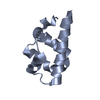
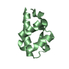
 Structure visualization
 Downloads & links
 Other downloads
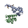


 PDBj
PDBj Assembly
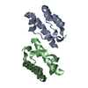

 Mass: 18.015 Da / Num. of mol.: 290 / Source method: isolated from a natural source / Formula: H2O
 Sample preparation
 / Beamline: 14-BM-C / Wavelength: 0.9002 Å
 Processing