 Movie
Movie Controller

[English] 日本語
 Yorodumi
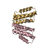
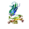
- PDB-5dfk: Crystal Structure of the Escherichia coli Common Pilus Chaperone, EcpB -
+ Open data

- Basic information
| Entry | Database: PDB / ID: 5dfk | ||||||
|---|---|---|---|---|---|---|---|
| Title | Crystal Structure of the Escherichia coli Common Pilus Chaperone, EcpB | ||||||
 Components Components | Probable fimbrial chaperone EcpB | ||||||
 Keywords Keywords | STRUCTURAL PROTEIN / chaperone usher / pilus / pili / biofilm / adhesion / biogenesis / E. coli / virulence | ||||||
| Function / homology | EcpB, C-terminal / EcpB C-terminal domain / PapD-like superfamily / Immunoglobulin-like fold / Probable fimbrial chaperone EcpB / Probable fimbrial chaperone EcpB Function and homology information Function and homology information | ||||||
| Biological species |  | ||||||
| Method |  X-RAY DIFFRACTION / SYNCHROTRON / SIRAS / Resolution: 2.4 Å X-RAY DIFFRACTION / SYNCHROTRON / SIRAS / Resolution: 2.4 Å | ||||||
 Authors Authors | Garnett, J.A. / Diallo, M. / Matthews, S.J. | ||||||
 Citation Citation | Journal: Plos Pathog. / Year: 2015 Title: Structural Insight into Archaic and Alternative Chaperone-Usher Pathways Reveals a Novel Mechanism of Pilus Biogenesis. Authors: Pakharukova, N. / Garnett, J.A. / Tuittila, M. / Paavilainen, S. / Diallo, M. / Xu, Y. / Matthews, S.J. / Zavialov, A.V. | ||||||
| History |
|
- Structure visualization
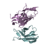
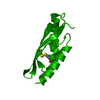
| Structure viewer | Molecule: MolmilJmol/JSmol |
|---|
- Downloads & links
-Download
| PDBx/mmCIF format | 5dfk.cif.gz | 87.5 KB | Display | PDBx/mmCIF format |
|---|---|---|---|---|
| PDB format | pdb5dfk.ent.gz | 64.6 KB | Display | PDB format |
| PDBx/mmJSON format | 5dfk.json.gz | Tree view | PDBx/mmJSON format | |
| Others |  Other downloads Other downloads |
-Validation report
| Arichive directory | https://data.pdbj.org/pub/pdb/validation_reports/df/5dfkftp://data.pdbj.org/pub/pdb/validation_reports/df/5dfk | HTTPS FTP |
|---|
-Related structure data


-Links
 PDBj
PDBj- Assembly
| Deposited unit | 
| ||||||||
|---|---|---|---|---|---|---|---|---|---|
| 1 |
| ||||||||
| Unit cell |
|
-Components
| #1: Protein | Mass: 24261.309 Da / Num. of mol.: 1 / Fragment: UNP residues 21-222 Source method: isolated from a genetically manipulated source Source: (gene. exp.) |
|---|---|
| #2: Water | ChemComp-HOH /  Mass: 18.015 Da / Num. of mol.: 73 / Source method: isolated from a natural source / Formula: H2O Mass: 18.015 Da / Num. of mol.: 73 / Source method: isolated from a natural source / Formula: H2O |
| Has protein modification | Y |
-Experimental details
-Experiment
| Experiment | Method: X-RAY DIFFRACTION |
|---|
- Sample preparation
| Crystal | Density Matthews: 2.19 Å3/Da / Density % sol: 43.86 % |
|---|---|
| Crystal grow | Temperature: 293 K / Method: vapor diffusion, sitting drop / Details: 15% (v/v) glycerol, 15% (w/v) PEG 5000 MME |
-Data collection
| Diffraction | Mean temperature: 100 K |
|---|---|
| Diffraction source | Source: SYNCHROTRON / Site: Diamond  / Beamline: I04 / Wavelength: 0.97949 Å / Beamline: I04 / Wavelength: 0.97949 Å |
| Detector | Type: DECTRIS PILATUS 6M / Detector: PIXEL / Date: Jul 7, 2013 |
| Radiation | Protocol: SINGLE WAVELENGTH / Monochromatic (M) / Laue (L): M / Scattering type: x-ray |
| Radiation wavelength | Wavelength: 0.97949 Å / Relative weight: 1 |
| Reflection | Resolution: 2.4→54.26 Å / Num. obs: 11265 / % possible obs: 99.9 % / Redundancy: 19.2 % / Biso Wilson estimate: 32.306 Å2 / Rmerge(I) obs: 0.057 / Net I/σ(I): 44.4 |
| Reflection shell | Resolution: 2.4→2.46 Å / Redundancy: 19.4 % / Rmerge(I) obs: 0.492 / Mean I/σ(I) obs: 6.6 / % possible all: 99.7 |
- Processing
| Software |
| ||||||||||||||||||||||||||||||||||||||||||||||||||||||||||||||||||||||||||||||||||||||||||||||||||||||||||||||||||||||||||||||||||||||||||||||||||||||||||||||||||||||||||||||||||||||
|---|---|---|---|---|---|---|---|---|---|---|---|---|---|---|---|---|---|---|---|---|---|---|---|---|---|---|---|---|---|---|---|---|---|---|---|---|---|---|---|---|---|---|---|---|---|---|---|---|---|---|---|---|---|---|---|---|---|---|---|---|---|---|---|---|---|---|---|---|---|---|---|---|---|---|---|---|---|---|---|---|---|---|---|---|---|---|---|---|---|---|---|---|---|---|---|---|---|---|---|---|---|---|---|---|---|---|---|---|---|---|---|---|---|---|---|---|---|---|---|---|---|---|---|---|---|---|---|---|---|---|---|---|---|---|---|---|---|---|---|---|---|---|---|---|---|---|---|---|---|---|---|---|---|---|---|---|---|---|---|---|---|---|---|---|---|---|---|---|---|---|---|---|---|---|---|---|---|---|---|---|---|---|---|
| Refinement | Method to determine structure: SIRAS / Resolution: 2.4→54.26 Å / Cor.coef. Fo:Fc: 0.94 / Cor.coef. Fo:Fc free: 0.925 / SU B: 13.018 / SU ML: 0.157 / Cross valid method: THROUGHOUT / ESU R: 0.282 / ESU R Free: 0.217 / Stereochemistry target values: MAXIMUM LIKELIHOOD / Details: HYDROGENS HAVE BEEN ADDED IN THE RIDING POSITIONS
| ||||||||||||||||||||||||||||||||||||||||||||||||||||||||||||||||||||||||||||||||||||||||||||||||||||||||||||||||||||||||||||||||||||||||||||||||||||||||||||||||||||||||||||||||||||||
| Solvent computation | Ion probe radii: 0.8 Å / Shrinkage radii: 0.8 Å / VDW probe radii: 1.2 Å / Solvent model: MASK | ||||||||||||||||||||||||||||||||||||||||||||||||||||||||||||||||||||||||||||||||||||||||||||||||||||||||||||||||||||||||||||||||||||||||||||||||||||||||||||||||||||||||||||||||||||||
| Displacement parameters | Biso mean: 42.69 Å2
| ||||||||||||||||||||||||||||||||||||||||||||||||||||||||||||||||||||||||||||||||||||||||||||||||||||||||||||||||||||||||||||||||||||||||||||||||||||||||||||||||||||||||||||||||||||||
| Refinement step | Cycle: 1 / Resolution: 2.4→54.26 Å
| ||||||||||||||||||||||||||||||||||||||||||||||||||||||||||||||||||||||||||||||||||||||||||||||||||||||||||||||||||||||||||||||||||||||||||||||||||||||||||||||||||||||||||||||||||||||
| Refine LS restraints |
|