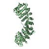
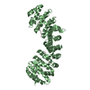
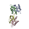
Co-inhibition by PD-1 / negative regulation of tolerance induction / negative regulation of immune response / negative regulation of T cell mediated immune response to tumor cell / positive regulation of T cell apoptotic process / negative regulation of interleukin-10 production / negative regulation of T cell activation / negative regulation of activated T cell proliferation / negative regulation of type II interferon production / negative regulation of T cell receptor signaling pathway ...Co-inhibition by PD-1 / negative regulation of tolerance induction / negative regulation of immune response / negative regulation of T cell mediated immune response to tumor cell / positive regulation of T cell apoptotic process / negative regulation of interleukin-10 production / negative regulation of T cell activation / negative regulation of activated T cell proliferation / negative regulation of type II interferon production / negative regulation of T cell receptor signaling pathway / regulation of immune response / negative regulation of T cell proliferation / T cell costimulation / positive regulation of T cell proliferation / transmembrane signaling receptor activity / cellular response to lipopolysaccharide / signaling receptor activity / adaptive immune response / cell surface receptor signaling pathway / immune response / positive regulation of apoptotic process / external side of plasma membrane / apoptotic process / negative regulation of apoptotic process / plasma membrane Similarity search - Function
Resolution: 1.6→23.98 Å / Cor.coef. Fo:Fc: 0.962 / Cor.coef. Fo:Fc free: 0.942 / SU B: 1.684 / SU ML: 0.06 / Cross valid method: THROUGHOUT / ESU R: 0.085 / ESU R Free: 0.088 / Stereochemistry target values: MAXIMUM LIKELIHOOD / Details: HYDROGENS HAVE BEEN ADDED IN THE RIDING POSITIONS
Rfactor
Num. reflection
% reflection
Selection details
Rfree
0.21833
2405
5 %
RANDOM
Rwork
0.18407
-
-
-
obs
0.18583
45379
99.59 %
-
all
-
47784
-
-
Solvent computation
Ion probe radii: 0.8 Å / Shrinkage radii: 0.8 Å / VDW probe radii: 1.2 Å / Solvent model: MASK
Displacement parameters
Biso mean: 25.773 Å2
Baniso -1
Baniso -2
Baniso -3
1-
0.01 Å2
0 Å2
0 Å2
2-
-
0 Å2
0 Å2
3-
-
-
-0.01 Å2
Refinement step
Cycle: LAST / Resolution: 1.6→23.98 Å
Protein
Nucleic acid
Ligand
Solvent
Total
Num. atoms
2347
0
24
278
2649
Refine LS restraints
Refine-ID
Type
Dev ideal
Dev ideal target
Number
X-RAY DIFFRACTION
r_bond_refined_d
0.008
0.022
2553
X-RAY DIFFRACTION
r_bond_other_d
X-RAY DIFFRACTION
r_angle_refined_deg
1.251
1.963
3494
X-RAY DIFFRACTION
r_angle_other_deg
X-RAY DIFFRACTION
r_dihedral_angle_1_deg
5.979
5
326
X-RAY DIFFRACTION
r_dihedral_angle_2_deg
36.193
24
110
X-RAY DIFFRACTION
r_dihedral_angle_3_deg
14.006
15
425
X-RAY DIFFRACTION
r_dihedral_angle_4_deg
21.055
15
17
X-RAY DIFFRACTION
r_chiral_restr
0.086
0.2
394
X-RAY DIFFRACTION
r_gen_planes_refined
0.004
0.02
1947
X-RAY DIFFRACTION
r_gen_planes_other
X-RAY DIFFRACTION
r_nbd_refined
0.178
0.3
1033
X-RAY DIFFRACTION
r_nbd_other
X-RAY DIFFRACTION
r_nbtor_refined
0.307
0.5
1717
X-RAY DIFFRACTION
r_nbtor_other
X-RAY DIFFRACTION
r_xyhbond_nbd_refined
0.15
0.5
458
X-RAY DIFFRACTION
r_xyhbond_nbd_other
X-RAY DIFFRACTION
r_metal_ion_refined
X-RAY DIFFRACTION
r_metal_ion_other
X-RAY DIFFRACTION
r_symmetry_vdw_refined
0.207
0.3
55
X-RAY DIFFRACTION
r_symmetry_vdw_other
X-RAY DIFFRACTION
r_symmetry_hbond_refined
0.147
0.5
32
X-RAY DIFFRACTION
r_symmetry_hbond_other
X-RAY DIFFRACTION
r_symmetry_metal_ion_refined
X-RAY DIFFRACTION
r_symmetry_metal_ion_other
X-RAY DIFFRACTION
r_mcbond_it
2.198
2
1617
X-RAY DIFFRACTION
r_mcbond_other
X-RAY DIFFRACTION
r_mcangle_it
3.326
3
2592
X-RAY DIFFRACTION
r_scbond_it
3.306
3
1053
X-RAY DIFFRACTION
r_scangle_it
5.179
5
897
X-RAY DIFFRACTION
r_rigid_bond_restr
X-RAY DIFFRACTION
r_sphericity_free
X-RAY DIFFRACTION
r_sphericity_bonded
LS refinement shell
Resolution: 1.6→1.637 Å / Total num. of bins used: 20
Rfactor
Num. reflection
% reflection
Rfree
0.288
169
-
Rwork
0.249
3280
-
obs
-
-
97.4 %
+
About Yorodumi
-
News
-
Feb 9, 2022. New format data for meta-information of EMDB entries
New format data for meta-information of EMDB entries
Version 3 of the EMDB header file is now the official format.
The previous official version 1.9 will be removed from the archive.
In the structure databanks used in Yorodumi, some data are registered as the other names, "COVID-19 virus" and "2019-nCoV". Here are the details of the virus and the list of structure data.
Jan 31, 2019. EMDB accession codes are about to change! (news from PDBe EMDB page)
EMDB accession codes are about to change! (news from PDBe EMDB page)
The allocation of 4 digits for EMDB accession codes will soon come to an end. Whilst these codes will remain in use, new EMDB accession codes will include an additional digit and will expand incrementally as the available range of codes is exhausted. The current 4-digit format prefixed with “EMD-” (i.e. EMD-XXXX) will advance to a 5-digit format (i.e. EMD-XXXXX), and so on. It is currently estimated that the 4-digit codes will be depleted around Spring 2019, at which point the 5-digit format will come into force.
The EM Navigator/Yorodumi systems omit the EMD- prefix.
Related info.:Q: What is EMD? / ID/Accession-code notation in Yorodumi/EM Navigator
Yorodumi is a browser for structure data from EMDB, PDB, SASBDB, etc.
This page is also the successor to EM Navigator detail page, and also detail information page/front-end page for Omokage search.
The word "yorodu" (or yorozu) is an old Japanese word meaning "ten thousand". "mi" (miru) is to see.
Related info.:EMDB / PDB / SASBDB / Comparison of 3 databanks / Yorodumi Search / Aug 31, 2016. New EM Navigator & Yorodumi / Yorodumi Papers / Jmol/JSmol / Function and homology information / Changes in new EM Navigator and Yorodumi
 Movie
Movie Controller


 Open data
 Basic information
 Components
 Keywords
 Function and homology information
 X-RAY DIFFRACTION /
  Authors
 Citation
 Structure visualization
 Downloads & links
 Other downloads

 PDBj
PDBj


 Assembly


 Mass: 92.094 Da / Num. of mol.: 4 / Source method: obtained synthetically / Formula: C3H8O3
 Mass: 18.015 Da / Num. of mol.: 278 / Source method: isolated from a natural source / Formula: H2O
 Sample preparation
 / Beamline: X29A / Wavelength: 0.9792 Å
 Processing