 Movie
Movie Controller

[English] 日本語
 Yorodumi
- PDB-3boc: Carbonic anhydrase from marine diatom Thalassiosira weissflogii- ... -
+ Open data

- Basic information
| Entry | Database: PDB / ID: 3boc | ||||||
|---|---|---|---|---|---|---|---|
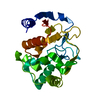
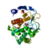
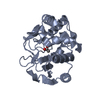
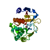
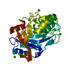
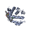
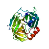
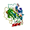
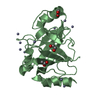
| Title | Carbonic anhydrase from marine diatom Thalassiosira weissflogii- zinc bound domain 2 (CDCA1-R2) | ||||||
 Components Components | Cadmium-specific carbonic anhydrase | ||||||
 Keywords Keywords | LYASE / carbonic anhydrase / marine diatom / zinc-bound | ||||||
| Function / homology | Cadmium carbonic anhydrase repeat / Cadmium carbonic anhydrase repeat / metal ion binding / Cadmium-specific carbonic anhydrase Function and homology information Function and homology information | ||||||
| Biological species |  Thalassiosira weissflogii (Diatom) Thalassiosira weissflogii (Diatom) | ||||||
| Method |  X-RAY DIFFRACTION / FOURIER SYNTHESIS / Resolution: 1.8 Å X-RAY DIFFRACTION / FOURIER SYNTHESIS / Resolution: 1.8 Å | ||||||
 Authors Authors | Xu, Y. / Feng, L. / Jeffrey, P.D. / Shi, Y. / Morel, F.M.M. | ||||||
 Citation Citation | Journal: Nature / Year: 2008 Title: Structure and metal exchange in the cadmium carbonic anhydrase of marine diatoms. Authors: Xu, Y. / Feng, L. / Jeffrey, P.D. / Shi, Y. / Morel, F.M. | ||||||
| History |
|
- Structure visualization
| Structure viewer | Molecule: MolmilJmol/JSmol |
|---|
- Downloads & links
-Download
| PDBx/mmCIF format | 3boc.cif.gz | 54.9 KB | Display | PDBx/mmCIF format |
|---|---|---|---|---|
| PDB format | pdb3boc.ent.gz | 38.5 KB | Display | PDB format |
| PDBx/mmJSON format | 3boc.json.gz | Tree view | PDBx/mmJSON format | |
| Others |  Other downloads Other downloads |
-Validation report
| Arichive directory | https://data.pdbj.org/pub/pdb/validation_reports/bo/3bocftp://data.pdbj.org/pub/pdb/validation_reports/bo/3boc | HTTPS FTP |
|---|
-Related structure data


-Links
 PDBj
PDBj
- Assembly
| Deposited unit | 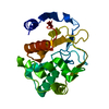
| ||||||||
|---|---|---|---|---|---|---|---|---|---|
| 1 |
| ||||||||
| Unit cell |
|
-Components
| #1: Protein | Mass: 22432.449 Da / Num. of mol.: 1 / Fragment: Domain 2, CDCA1-R2 Source method: isolated from a genetically manipulated source Source: (gene. exp.) Thalassiosira weissflogii (Diatom) / Gene: cdca1 / Production host:  |
|---|---|
| #2: Chemical | ChemComp-ZN /   Mass: 65.409 Da / Num. of mol.: 1 / Source method: obtained synthetically / Formula: Zn Mass: 65.409 Da / Num. of mol.: 1 / Source method: obtained synthetically / Formula: Zn |
| #3: Water | ChemComp-HOH /  Mass: 18.015 Da / Num. of mol.: 127 / Source method: isolated from a natural source / Formula: H2O Mass: 18.015 Da / Num. of mol.: 127 / Source method: isolated from a natural source / Formula: H2O |
| Nonpolymer details | RELATIVE TO THE ORIGINAL COORDINATES THE ZINC AND WATER MOLECULE NUMBERING HAS BEEN OFFSET BY 1000, ...RELATIVE TO THE ORIGINAL COORDINATE |
-Experimental details
-Experiment
| Experiment | Method: X-RAY DIFFRACTION |
|---|
- Sample preparation
| Crystal | Density Matthews: 2.12 Å3/Da / Density % sol: 42.03 % |
|---|---|
| Crystal grow | Temperature: 298 K / Method: vapor diffusion, hanging drop / pH: 5.5 Details: 0.1 M sodium citrate, 20% (w/v) PEG3350, pH 5.5, VAPOR DIFFUSION, HANGING DROP, temperature 298K |
-Data collection
| Diffraction | Mean temperature: 100 K |
|---|---|
| Diffraction source | Source: ROTATING ANODE / Type: RIGAKU RU300 / Wavelength: 1.54 Å |
| Detector | Type: RIGAKU RAXIS IV++ / Detector: IMAGE PLATE / Date: Dec 4, 2006 |
| Radiation | Monochromator: Mirrors / Protocol: SINGLE WAVELENGTH / Monochromatic (M) / Laue (L): M / Scattering type: x-ray |
| Radiation wavelength | Wavelength: 1.54 Å / Relative weight: 1 |
| Reflection | Resolution: 1.8→100 Å / Num. all: 17726 / Num. obs: 17726 / % possible obs: 96.4 % / Observed criterion σ(I): -3 / Redundancy: 7.3 % / Biso Wilson estimate: 23.4 Å2 / Rsym value: 0.054 |
| Reflection shell | Resolution: 1.8→1.86 Å / Rsym value: 0.284 / % possible all: 88 |
- Processing
| Software |
| ||||||||||||||||||||||||||||||||||||||||||||||||||||||||||||||||||||||||||||||||
|---|---|---|---|---|---|---|---|---|---|---|---|---|---|---|---|---|---|---|---|---|---|---|---|---|---|---|---|---|---|---|---|---|---|---|---|---|---|---|---|---|---|---|---|---|---|---|---|---|---|---|---|---|---|---|---|---|---|---|---|---|---|---|---|---|---|---|---|---|---|---|---|---|---|---|---|---|---|---|---|---|---|
| Refinement | Method to determine structure: FOURIER SYNTHESIS / Resolution: 1.8→25 Å / Rfactor Rfree error: 0.009 / Data cutoff high absF: 1089886.45 / Data cutoff low absF: 0 / Isotropic thermal model: RESTRAINED / Cross valid method: THROUGHOUT / σ(F): 0 / Stereochemistry target values: Engh & Huber
| ||||||||||||||||||||||||||||||||||||||||||||||||||||||||||||||||||||||||||||||||
| Solvent computation | Solvent model: FLAT MODEL / Bsol: 49.9538 Å2 / ksol: 0.335251 e/Å3 | ||||||||||||||||||||||||||||||||||||||||||||||||||||||||||||||||||||||||||||||||
| Displacement parameters | Biso mean: 28.7 Å2
| ||||||||||||||||||||||||||||||||||||||||||||||||||||||||||||||||||||||||||||||||
| Refine analyze |
| ||||||||||||||||||||||||||||||||||||||||||||||||||||||||||||||||||||||||||||||||
| Refinement step | Cycle: LAST / Resolution: 1.8→25 Å
| ||||||||||||||||||||||||||||||||||||||||||||||||||||||||||||||||||||||||||||||||
| Refine LS restraints |
| ||||||||||||||||||||||||||||||||||||||||||||||||||||||||||||||||||||||||||||||||
| LS refinement shell | Resolution: 1.8→1.91 Å / Rfactor Rfree error: 0.034 / Total num. of bins used: 6
| ||||||||||||||||||||||||||||||||||||||||||||||||||||||||||||||||||||||||||||||||
| Xplor file |
|
