SEQUENCE THE CONSTRUCT WAS EXPRESSED WITH A PURIFICATION TAG MGSDKIHHHHHHENLYFQG. THE CONSTRUCT ... SEQUENCE THE CONSTRUCT WAS EXPRESSED WITH A PURIFICATION TAG MGSDKIHHHHHHENLYFQG. THE CONSTRUCT WAS ENGINEERED WITH THE FOLLOWING MUTATIONS: E112A, K113A, K115A.
Mass: 18.015 Da / Num. of mol.: 214 / Source method: isolated from a natural source / Formula: H2O
Has protein modification
Y
Sequence details
REMARK 999 SEQUENCE: THE CONSTRUCT WAS EXPRESSED WITH A PURIFICATION REMARK 999 TAG ...REMARK 999 SEQUENCE: THE CONSTRUCT WAS EXPRESSED WITH A PURIFICATION REMARK 999 TAG MGSDKIHHHHHHENLYFQG. THE CONSTRUCT WAS ENGINEERED WITH REMARK 999 THE FOLLOWING MUTATIONS: E112A, K113A, K115A.
-
Experimental details
-
Experiment
Experiment
Method: X-RAY DIFFRACTION / Number of used crystals: 1
-
Sample preparation
Crystal
Density Matthews: 2.08 Å3/Da / Density % sol: 40.9 %
Crystal grow
Temperature: 277 K / Method: vapor diffusion, sitting drop / pH: 4.5 Details: NANODROP, 40.0% 1,2-propanediol, 0.1M Acetate pH 4.5, VAPOR DIFFUSION, SITTING DROP, temperature 277K
Resolution: 1.91→29.185 Å / Num. obs: 46740 / % possible obs: 98.4 % / Observed criterion σ(I): -3 / Biso Wilson estimate: 36.803 Å2 / Rmerge(I) obs: 0.05 / Net I/σ(I): 10.78
Reflection shell
Resolution (Å)
Rmerge(I) obs
Mean I/σ(I) obs
Num. measured obs
Num. unique obs
Diffraction-ID
% possible all
1.91-1.98
0.685
1.4
14080
9105
1
99.6
1.98-2.06
0.527
1.8
13852
8909
1
99.6
2.06-2.15
0.396
2.4
13398
8550
1
99.5
2.15-2.26
0.293
3.2
13680
8669
1
99.5
2.26-2.41
0.219
4.2
14785
9353
1
99.6
2.41-2.59
0.153
5.9
13789
8585
1
99.3
2.59-2.85
0.094
9
14335
8825
1
98.8
2.85-3.26
0.046
16
14478
8767
1
98.1
3.26-4.1
0.024
27.8
14586
8704
1
97.6
4.1-29.185
0.017
37.8
14360
8401
1
92.8
-
Phasing
Phasing
Method: MAD
-
Processing
Software
Name
Version
Classification
NB
REFMAC
5.2.0019
refinement
PHENIX
refinement
SOLVE
phasing
MolProbity
3beta29
modelbuilding
XSCALE
datascaling
PDB_EXTRACT
3
dataextraction
MAR345
CCD
datacollection
XDS
datareduction
Refinement
Method to determine structure: MAD / Resolution: 1.91→29.185 Å / Cor.coef. Fo:Fc: 0.958 / Cor.coef. Fo:Fc free: 0.94 / SU B: 10.13 / SU ML: 0.137 / TLS residual ADP flag: LIKELY RESIDUAL / Cross valid method: THROUGHOUT / σ(F): 0 / ESU R: 0.168 / ESU R Free: 0.155 Stereochemistry target values: MAXIMUM LIKELIHOOD WITH PHASES Details: 1. HYDROGENS HAVE BEEN ADDED IN THE RIDING POSITIONS. 2. ATOM RECORDS CONTAIN RESIDUAL B FACTORS ONLY. 3. A MET-INHIBITION PROTOCOL WAS USED FOR SELENOMETHIONINE INCORPORATION DURING PROTEIN ...Details: 1. HYDROGENS HAVE BEEN ADDED IN THE RIDING POSITIONS. 2. ATOM RECORDS CONTAIN RESIDUAL B FACTORS ONLY. 3. A MET-INHIBITION PROTOCOL WAS USED FOR SELENOMETHIONINE INCORPORATION DURING PROTEIN EXPRESSION. THE OCCUPANCY OF THE SE ATOMS IN THE MSE RESIDUES WAS REDUCED TO 0.75 FOR THE REDUCED SCATTERING POWER DUE TO PARTIAL S-MET INCORPORATION. 4. 1,2-PROPANEDIOL (PGO) IS MODELED FROM THE CRYSTALLIZATION CONDITION.
Rfactor
Num. reflection
% reflection
Selection details
Rfree
0.245
2364
5.1 %
RANDOM
Rwork
0.202
-
-
-
obs
0.205
46708
99.12 %
-
Solvent computation
Ion probe radii: 0.8 Å / Shrinkage radii: 0.8 Å / VDW probe radii: 1.2 Å / Solvent model: BABINET MODEL WITH MASK
In the structure databanks used in Yorodumi, some data are registered as the other names, "COVID-19 virus" and "2019-nCoV". Here are the details of the virus and the list of structure data.
Jan 31, 2019. EMDB accession codes are about to change! (news from PDBe EMDB page)
EMDB accession codes are about to change! (news from PDBe EMDB page)
The allocation of 4 digits for EMDB accession codes will soon come to an end. Whilst these codes will remain in use, new EMDB accession codes will include an additional digit and will expand incrementally as the available range of codes is exhausted. The current 4-digit format prefixed with “EMD-” (i.e. EMD-XXXX) will advance to a 5-digit format (i.e. EMD-XXXXX), and so on. It is currently estimated that the 4-digit codes will be depleted around Spring 2019, at which point the 5-digit format will come into force.
The EM Navigator/Yorodumi systems omit the EMD- prefix.
Related info.:Q: What is EMD? / ID/Accession-code notation in Yorodumi/EM Navigator
Yorodumi is a browser for structure data from EMDB, PDB, SASBDB, etc.
This page is also the successor to EM Navigator detail page, and also detail information page/front-end page for Omokage search.
The word "yorodu" (or yorozu) is an old Japanese word meaning "ten thousand". "mi" (miru) is to see.
Related info.:EMDB / PDB / SASBDB / Comparison of 3 databanks / Yorodumi Search / Aug 31, 2016. New EM Navigator & Yorodumi / Yorodumi Papers / Jmol/JSmol / Function and homology information / Changes in new EM Navigator and Yorodumi
 Movie
Movie Controller

 Yorodumi
 Open data
 Basic information
 Components
 Keywords
 Function and homology information
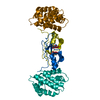
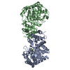
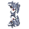
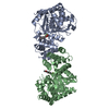
 Thermoplasma acidophilum DSM 1728 (acidophilic)
 X-RAY DIFFRACTION /
  Authors
 Citation
 Structure visualization
 Downloads & links
 Other downloads


 PDBj
PDBj Assembly


 Mass: 76.094 Da / Num. of mol.: 7 / Source method: obtained synthetically / Formula: C3H8O2
 Mass: 94.971 Da / Num. of mol.: 1 / Source method: obtained synthetically / Formula: PO4
 Mass: 18.015 Da / Num. of mol.: 214 / Source method: isolated from a natural source / Formula: H2O
 Sample preparation
 / Beamline: 23-ID-D / Wavelength: 0.91840, 0.97939, 0.97953
 Processing