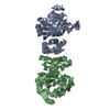
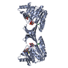
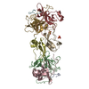
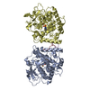
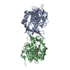
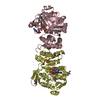
BIOMOLECULE: 1,2 THIS ENTRY CONTAINS THE CRYSTALLOGRAPHIC ASYMMETRIC UNIT WHICH CONSISTS OF 4 ...BIOMOLECULE: 1,2 THIS ENTRY CONTAINS THE CRYSTALLOGRAPHIC ASYMMETRIC UNIT WHICH CONSISTS OF 4 CHAIN(S). SEE REMARK 350 FOR INFORMATION ON GENERATING THE BIOLOGICAL MOLECULE(S). SIZE EXCLUSION CHROMATOGRAPHY SUPPORTS THE ASSIGNMENT OF A DIMER AS A BIOLOGICALLY SIGNIFICANT OLIGOMERIZATION STATE.
Remark 999
SEQUENCE THE CONSTRUCT WAS EXPRESSED WITH A PURIFICATION TAG MGSDKIHHHHHHENLYFQG. THE TAG WAS ...SEQUENCE THE CONSTRUCT WAS EXPRESSED WITH A PURIFICATION TAG MGSDKIHHHHHHENLYFQG. THE TAG WAS REMOVED WITH TEV PROTEASE LEAVING ONLY A GLYCINE (0) FOLLOWED BY THE TARGET SEQUENCE. DNA SEQUENCING INDICATES THAT RESIDUE 7 IS ISOLEUCINE (NOT VALINE) IN THE CLONED CONSTRUCT. THE SEQUENCING RESULTS ARE CONSISTENT WITH THE ELECTRON DENSITY AND MASS SPECTROMETRY RESULTS FOR THE EXPRESSED PROTEIN.
Monochromator: Double Crystal Si(111) / Protocol: SINGLE WAVELENGTH / Monochromatic (M) / Laue (L): M / Scattering type: x-ray
Radiation wavelength
Wavelength: 0.9797 Å / Relative weight: 1
Reflection
Resolution: 2.6→29.437 Å / Num. obs: 54381 / % possible obs: 99.6 % / Biso Wilson estimate: 45.6 Å2 / Rmerge(I) obs: 0.083 / Net I/σ(I): 15.41
Reflection shell
Resolution (Å)
Rmerge(I) obs
Mean I/σ(I) obs
Num. measured obs
% possible all
2.6-2.69
0.401
2.9
33997
97.5
2.69-2.8
0.339
3.7
40153
100
2.8-2.93
0.273
4.7
40574
100
2.93-3.08
0.196
6.9
38502
100
3.08-3.27
0.144
9.3
39080
100
3.27-3.52
0.095
13.7
39277
99.9
3.52-3.88
0.06
20.8
40256
100
3.88-4.43
0.046
26.6
38861
99.9
4.43-5.57
0.043
29.7
39206
99.9
5.57-29.4
0.029
35
39032
98.8
-
Phasing
Phasing
Method: SAD
-
Processing
Software
Name
Version
Classification
NB
MolProbity
3beta29
modelbuilding
SHELX
phasing
REFMAC
5.2.0019
refinement
XSCALE
datascaling
PDB_EXTRACT
2
dataextraction
XDS
datareduction
SHELXD
phasing
autoSHARP
phasing
Refinement
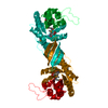
Method to determine structure: SAD / Resolution: 2.6→29.437 Å / Cor.coef. Fo:Fc: 0.937 / Cor.coef. Fo:Fc free: 0.913 / SU B: 16.706 / SU ML: 0.176 / Cross valid method: THROUGHOUT / σ(F): 0 / ESU R: 0.425 / ESU R Free: 0.255 Stereochemistry target values: MAXIMUM LIKELIHOOD WITH PHASES Details: 1. HYDROGENS HAVE BEEN ADDED IN THE RIDING POSITIONS. 2. ONE NAD IS ASSOCIATED WITH EACH MONOMER. NAD IS ASSIGNED BASED ON ELECTRON DENSITY. THE ASSIGNMENT IS TENTATIVE DUE TO POOR DENSITY ...Details: 1. HYDROGENS HAVE BEEN ADDED IN THE RIDING POSITIONS. 2. ONE NAD IS ASSOCIATED WITH EACH MONOMER. NAD IS ASSIGNED BASED ON ELECTRON DENSITY. THE ASSIGNMENT IS TENTATIVE DUE TO POOR DENSITY AND LIMITED RESOLUTION, ONLY PYROPHOSPHATE AND RIBOSE DENSITY ARE DEFINITIVE. 3. A MET-INHIBITION PROTOCOL WAS USED FOR SELENOMETHIONINE INCORPOR DURING PROTEIN EXPRESSION. THE OCCUPANCY OF THE SE ATOMS IN THE MSE RESIDUES WAS REDUCED TO 0.7 TO ACCOUNT FOR THE REDUCED SCATTERING DUE TO PARTIAL S-MET INCORPORATION.
Rfactor
Num. reflection
% reflection
Selection details
Rfree
0.224
2764
5.1 %
RANDOM
Rwork
0.188
-
-
-
obs
0.19
54312
99.68 %
-
Solvent computation
Ion probe radii: 0.8 Å / Shrinkage radii: 0.8 Å / VDW probe radii: 1.2 Å / Solvent model: BABINET MODEL WITH MASK
Displacement parameters
Biso mean: 36.271 Å2
Baniso -1
Baniso -2
Baniso -3
1-
-0.86 Å2
0 Å2
0 Å2
2-
-
0.2 Å2
0 Å2
3-
-
-
0.66 Å2
Refinement step
Cycle: LAST / Resolution: 2.6→29.437 Å
Protein
Nucleic acid
Ligand
Solvent
Total
Num. atoms
9095
0
216
280
9591
Refine LS restraints
Refine-ID
Type
Dev ideal
Dev ideal target
Number
X-RAY DIFFRACTION
r_bond_refined_d
0.014
0.022
9442
X-RAY DIFFRACTION
r_bond_other_d
0.002
0.02
6217
X-RAY DIFFRACTION
r_angle_refined_deg
1.5
2.018
12822
X-RAY DIFFRACTION
r_angle_other_deg
0.92
3
15382
X-RAY DIFFRACTION
r_dihedral_angle_1_deg
5.492
5
1226
X-RAY DIFFRACTION
r_dihedral_angle_2_deg
35.56
24.936
312
X-RAY DIFFRACTION
r_dihedral_angle_3_deg
15.916
15
1618
X-RAY DIFFRACTION
r_dihedral_angle_4_deg
18.814
15
42
X-RAY DIFFRACTION
r_chiral_restr
0.072
0.2
1559
X-RAY DIFFRACTION
r_gen_planes_refined
0.004
0.02
10234
X-RAY DIFFRACTION
r_gen_planes_other
0.002
0.02
1610
X-RAY DIFFRACTION
r_nbd_refined
0.216
0.2
1874
X-RAY DIFFRACTION
r_nbd_other
0.188
0.2
6251
X-RAY DIFFRACTION
r_nbtor_refined
0.175
0.2
4557
X-RAY DIFFRACTION
r_nbtor_other
0.086
0.2
5055
X-RAY DIFFRACTION
r_xyhbond_nbd_refined
0.17
0.2
340
X-RAY DIFFRACTION
r_xyhbond_nbd_other
0.081
0.2
2
X-RAY DIFFRACTION
r_symmetry_vdw_refined
0.342
0.2
10
X-RAY DIFFRACTION
r_symmetry_vdw_other
0.174
0.2
42
X-RAY DIFFRACTION
r_symmetry_hbond_refined
0.098
0.2
4
X-RAY DIFFRACTION
r_mcbond_it
1.566
3
6313
X-RAY DIFFRACTION
r_mcbond_other
0.238
3
2543
X-RAY DIFFRACTION
r_mcangle_it
2.6
5
9827
X-RAY DIFFRACTION
r_scbond_it
4.899
8
3518
X-RAY DIFFRACTION
r_scangle_it
6.797
11
2995
Refine LS restraints NCS
Ens-ID: 1 / Refine-ID: X-RAY DIFFRACTION
Dom-ID
Auth asym-ID
Number
Type
Rms dev position (Å)
Weight position
1
A
1766
TIGHTPOSITIONAL
0.04
0.05
2
B
1766
TIGHTPOSITIONAL
0.04
0.05
3
C
1766
TIGHTPOSITIONAL
0.04
0.05
4
D
1766
TIGHTPOSITIONAL
0.04
0.05
1
A
1869
MEDIUMPOSITIONAL
0.34
0.5
2
B
1869
MEDIUMPOSITIONAL
0.3
0.5
3
C
1869
MEDIUMPOSITIONAL
0.28
0.5
4
D
1869
MEDIUMPOSITIONAL
0.29
0.5
1
A
6
LOOSEPOSITIONAL
0.13
5
2
B
6
LOOSEPOSITIONAL
0.33
5
3
C
6
LOOSEPOSITIONAL
0.38
5
4
D
6
LOOSEPOSITIONAL
0.62
5
1
A
1766
TIGHTTHERMAL
0.11
0.5
2
B
1766
TIGHTTHERMAL
0.11
0.5
3
C
1766
TIGHTTHERMAL
0.09
0.5
4
D
1766
TIGHTTHERMAL
0.1
0.5
1
A
1869
MEDIUMTHERMAL
0.88
2
2
B
1869
MEDIUMTHERMAL
0.78
2
3
C
1869
MEDIUMTHERMAL
0.7
2
4
D
1869
MEDIUMTHERMAL
0.73
2
1
A
6
LOOSETHERMAL
0.97
10
2
B
6
LOOSETHERMAL
1.42
10
3
C
6
LOOSETHERMAL
1.24
10
4
D
6
LOOSETHERMAL
1.29
10
LS refinement shell
Resolution: 2.599→2.667 Å / Total num. of bins used: 20
Rfactor
Num. reflection
% reflection
Rfree
0.299
205
-
Rwork
0.235
3654
-
obs
-
3859
97.8 %
+
About Yorodumi
-
News
-
Feb 9, 2022. New format data for meta-information of EMDB entries
New format data for meta-information of EMDB entries
Version 3 of the EMDB header file is now the official format.
The previous official version 1.9 will be removed from the archive.
In the structure databanks used in Yorodumi, some data are registered as the other names, "COVID-19 virus" and "2019-nCoV". Here are the details of the virus and the list of structure data.
Jan 31, 2019. EMDB accession codes are about to change! (news from PDBe EMDB page)
EMDB accession codes are about to change! (news from PDBe EMDB page)
The allocation of 4 digits for EMDB accession codes will soon come to an end. Whilst these codes will remain in use, new EMDB accession codes will include an additional digit and will expand incrementally as the available range of codes is exhausted. The current 4-digit format prefixed with “EMD-” (i.e. EMD-XXXX) will advance to a 5-digit format (i.e. EMD-XXXXX), and so on. It is currently estimated that the 4-digit codes will be depleted around Spring 2019, at which point the 5-digit format will come into force.
The EM Navigator/Yorodumi systems omit the EMD- prefix.
Related info.:Q: What is EMD? / ID/Accession-code notation in Yorodumi/EM Navigator
Yorodumi is a browser for structure data from EMDB, PDB, SASBDB, etc.
This page is also the successor to EM Navigator detail page, and also detail information page/front-end page for Omokage search.
The word "yorodu" (or yorozu) is an old Japanese word meaning "ten thousand". "mi" (miru) is to see.
Related info.:EMDB / PDB / SASBDB / Comparison of 3 databanks / Yorodumi Search / Aug 31, 2016. New EM Navigator & Yorodumi / Yorodumi Papers / Jmol/JSmol / Function and homology information / Changes in new EM Navigator and Yorodumi
 Movie
Movie Controller

 Yorodumi
 Open data
 Basic information
 Components
 Keywords
 Function and homology information
 Bacillus halodurans (bacteria)
 X-RAY DIFFRACTION /
  Authors
 Citation
 Structure visualization
 Downloads & links
 Other downloads
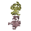


 PDBj
PDBj Assembly


 Mass: 96.063 Da / Num. of mol.: 8 / Source method: obtained synthetically / Formula: SO4
 Mass: 663.425 Da / Num. of mol.: 4 / Source method: obtained synthetically / Formula: C21H27N7O14P2 / Comment: NAD*YM
 Mass: 18.015 Da / Num. of mol.: 280 / Source method: isolated from a natural source / Formula: H2O
 Sample preparation
 / Beamline: 8.2.2 / Wavelength: 0.9797
  Processing