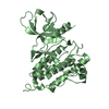
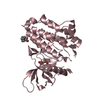
[English] 日本語
Yorodumi
- PDB-3bce: Crystal structure of the ErbB4 kinase -

+
Open data


ID or keywords:

Loading...

-
Basic information

Entry
Database: PDB / ID: 3bce
TitleCrystal structure of the ErbB4 kinase
ComponentsReceptor tyrosine-protein kinase erbB-4
KeywordsTRANSFERASE / active conformation / ATP-binding / Glycoprotein / Kinase / Membrane / Nucleotide-binding / Phosphorylation / Receptor / Transmembrane / Tyrosine-protein kinase
Function / homology
Function and homology information


establishment of planar polarity involved in nephron morphogenesis / ERBB4 signaling pathway / ERBB4-ERBB4 signaling pathway / olfactory bulb interneuron differentiation / central nervous system morphogenesis / neuregulin receptor activity / cardiac muscle tissue regeneration / ERBB2-ERBB4 signaling pathway / mitochondrial fragmentation involved in apoptotic process / GABA receptor binding ...establishment of planar polarity involved in nephron morphogenesis / ERBB4 signaling pathway / ERBB4-ERBB4 signaling pathway / olfactory bulb interneuron differentiation / central nervous system morphogenesis / neuregulin receptor activity / cardiac muscle tissue regeneration / ERBB2-ERBB4 signaling pathway / mitochondrial fragmentation involved in apoptotic process / GABA receptor binding / PI3K events in ERBB4 signaling / mammary gland epithelial cell differentiation / embryonic pattern specification / positive regulation of protein localization to cell surface / neurotransmitter receptor localization to postsynaptic specialization membrane / neural crest cell migration / epidermal growth factor receptor activity / epidermal growth factor receptor binding / ERBB2 Activates PTK6 Signaling / Signaling by ERBB4 / ERBB2 Regulates Cell Motility / cell surface receptor signaling pathway via JAK-STAT / Long-term potentiation / PI3K events in ERBB2 signaling / SHC1 events in ERBB4 signaling / GABA-ergic synapse / mammary gland alveolus development / cell fate commitment / Nuclear signaling by ERBB4 / positive regulation of tyrosine phosphorylation of STAT protein / positive regulation of cardiac muscle cell proliferation / regulation of cell migration / Signaling by ERBB2 / cellular response to epidermal growth factor stimulus / synapse assembly / cell surface receptor protein tyrosine kinase signaling pathway / Downregulation of ERBB4 signaling / lactation / GRB2 events in ERBB2 signaling / transmembrane receptor protein tyrosine kinase activity / SHC1 events in ERBB2 signaling / basal plasma membrane / neurogenesis / postsynaptic density membrane / Signaling by ERBB2 TMD/JMD mutants / positive regulation of receptor signaling pathway via JAK-STAT / neuromuscular junction / Signaling by ERBB2 KD Mutants / receptor protein-tyrosine kinase / Downregulation of ERBB2 signaling / peptidyl-tyrosine phosphorylation / Constitutive Signaling by Aberrant PI3K in Cancer / cell migration / PIP3 activates AKT signaling / presynaptic membrane / nervous system development / heart development / PI5P, PP2A and IER3 Regulate PI3K/AKT Signaling / RAF/MAP kinase cascade / basolateral plasma membrane / postsynaptic membrane / protein tyrosine kinase activity / Estrogen-dependent gene expression / protein autophosphorylation / positive regulation of phosphatidylinositol 3-kinase/protein kinase B signal transduction / positive regulation of ERK1 and ERK2 cascade / receptor complex / transcription cis-regulatory region binding / mitochondrial matrix / positive regulation of protein phosphorylation / negative regulation of cell population proliferation / glutamatergic synapse / positive regulation of cell population proliferation / negative regulation of apoptotic process / positive regulation of DNA-templated transcription / signal transduction / protein homodimerization activity / mitochondrion / extracellular region / nucleoplasm / ATP binding / nucleus / plasma membrane / cytosol
Similarity search - Function
: / Epidermal growth factor receptor transmembrane-juxtamembrane segment / Tyrosine protein kinase, EGF/ERB/XmrK receptor / Growth factor receptor domain 4 / Growth factor receptor domain IV / Receptor L-domain / Furin-like cysteine-rich domain / Receptor L-domain superfamily / Furin-like cysteine rich region / Receptor L domain ...: / Epidermal growth factor receptor transmembrane-juxtamembrane segment / Tyrosine protein kinase, EGF/ERB/XmrK receptor / Growth factor receptor domain 4 / Growth factor receptor domain IV / Receptor L-domain / Furin-like cysteine-rich domain / Receptor L-domain superfamily / Furin-like cysteine rich region / Receptor L domain / Furin-like repeat / Furin-like repeats / Growth factor receptor cysteine-rich domain superfamily / Tyrosine-protein kinase, catalytic domain / Tyrosine kinase, catalytic domain / Tyrosine protein kinases specific active-site signature. / Tyrosine-protein kinase, active site / Protein tyrosine and serine/threonine kinase / Serine-threonine/tyrosine-protein kinase, catalytic domain / Transferase(Phosphotransferase) domain 1 / Transferase(Phosphotransferase); domain 1 / Phosphorylase Kinase; domain 1 / Phosphorylase Kinase; domain 1 / Protein kinase, ATP binding site / Protein kinases ATP-binding region signature. / Protein kinase domain profile. / Protein kinase domain / Protein kinase-like domain superfamily / 2-Layer Sandwich / Orthogonal Bundle / Mainly Alpha / Alpha Beta
Similarity search - Domain/homology
: / DI(HYDROXYETHYL)ETHER / Receptor tyrosine-protein kinase erbB-4
Similarity search - Component
Biological speciesHomo sapiens (human)
MethodX-RAY DIFFRACTION / SYNCHROTRON / Resolution: 2.5 Å
AuthorsQiu, C.
CitationJournal: Structure / Year: 2008
Title: Mechanism of Activation and Inhibition of the HER4/ErbB4 Kinase.
Authors: Qiu, C. / Tarrant, M.K. / Choi, S.H. / Sathyamurthy, A. / Bose, R. / Banjade, S. / Pal, A. / Bornmann, W.G. / Lemmon, M.A. / Cole, P.A. / Leahy, D.J.
History
DepositionNov 12, 2007Deposition site: RCSB / Processing site: RCSB
Revision 1.0Feb 12, 2008Provider: repository / Type: Initial release
Revision 1.1Jul 13, 2011Group: Advisory / Version format compliance
Revision 1.2Oct 25, 2017Group: Advisory / Refinement description / Category: pdbx_unobs_or_zero_occ_atoms / software
Revision 1.3Feb 21, 2024Group: Advisory / Data collection ...Advisory / Data collection / Database references / Derived calculations / Refinement description
Category: chem_comp_atom / chem_comp_bond ...chem_comp_atom / chem_comp_bond / database_2 / pdbx_unobs_or_zero_occ_atoms / struct_ncs_dom_lim / struct_site
Item: _database_2.pdbx_DOI / _database_2.pdbx_database_accession ..._database_2.pdbx_DOI / _database_2.pdbx_database_accession / _struct_ncs_dom_lim.beg_auth_comp_id / _struct_ncs_dom_lim.end_auth_comp_id / _struct_site.pdbx_auth_asym_id / _struct_site.pdbx_auth_comp_id / _struct_site.pdbx_auth_seq_id

-
Structure visualization

Structure viewerMolecule:
MolmilJmol/JSmol

Downloads & links

-
Assembly

Deposited unit
A: Receptor tyrosine-protein kinase erbB-4
B: Receptor tyrosine-protein kinase erbB-4
C: Receptor tyrosine-protein kinase erbB-4
hetero molecules


Theoretical massNumber of molelcules
Total (without water)112,8306
Polymers112,4743
Non-polymers3553
Water3,891216
1
A: Receptor tyrosine-protein kinase erbB-4
hetero molecules


Theoretical massNumber of molelcules
Total (without water)37,5462
Polymers37,4911
Non-polymers551
Water181
TypeNameSymmetry operationNumber
identity operation1_555x,y,z1
MethodPISA
2
B: Receptor tyrosine-protein kinase erbB-4


Theoretical massNumber of molelcules
Total (without water)37,4911
Polymers37,4911
Non-polymers00
Water181
TypeNameSymmetry operationNumber
identity operation1_555x,y,z1
MethodPISA
3
C: Receptor tyrosine-protein kinase erbB-4
hetero molecules


Theoretical massNumber of molelcules
Total (without water)37,7923
Polymers37,4911
Non-polymers3002
Water181
TypeNameSymmetry operationNumber
identity operation1_555x,y,z1
MethodPISA
Unit cell
Length a, b, c (Å)86.723, 86.723, 120.008
Angle α, β, γ (deg.)90.000, 90.000, 120.000
Int Tables number145
Space group name H-MP32
Noncrystallographic symmetry (NCS)NCS domain:
IDEns-IDDetails
11A
21B
31C

NCS domain segments:

Component-ID: 1 / Ens-ID: 1 / Beg auth comp-ID: THR / Beg label comp-ID: THR / End auth comp-ID: ASP / End label comp-ID: ASP / Refine code: 4 / Auth seq-ID: 678 - 965 / Label seq-ID: 2 - 289

Dom-IDAuth asym-IDLabel asym-ID
1AA
2BB
3CC

-
Components

#1: Protein Receptor tyrosine-protein kinase erbB-4 / p180erbB4 / Tyrosine kinase-type cell surface receptor HER4


Mass: 37491.457 Da / Num. of mol.: 3 / Fragment: ErbB4 kinase domain (UNP residues 702-1029)
Source method: isolated from a genetically manipulated source
Source: (gene. exp.) Homo sapiens (human) / Gene: ERBB4, HER4 / Plasmid: pFastBac / Production host: Spodoptera frugiperda (fall armyworm) / Strain (production host): Sf9
References: UniProt: Q15303, receptor protein-tyrosine kinase
#2: Chemical ChemComp-MN / MANGANESE (II) ION


Mass: 54.938 Da / Num. of mol.: 1 / Source method: obtained synthetically / Formula: Mn
#3: Chemical ChemComp-PG4 / TETRAETHYLENE GLYCOL


Mass: 194.226 Da / Num. of mol.: 1 / Source method: obtained synthetically / Formula: C8H18O5 / Comment: precipitant*YM
#4: Chemical ChemComp-PEG / DI(HYDROXYETHYL)ETHER


Mass: 106.120 Da / Num. of mol.: 1 / Source method: obtained synthetically / Formula: C4H10O3
#5: Water ChemComp-HOH / water


Mass: 18.015 Da / Num. of mol.: 216 / Source method: isolated from a natural source / Formula: H2O

-
Experimental details

-
Experiment

ExperimentMethod: X-RAY DIFFRACTION / Number of used crystals: 1

-
Sample preparation

CrystalDensity Matthews: 2.32 Å3/Da / Density % sol: 46.9 %
Crystal growTemperature: 293 K / Method: vapor diffusion, hanging drop / pH: 8.5
Details: 15% PEG3350, 0.1 M Tris, pH 8.5, VAPOR DIFFUSION, HANGING DROP, temperature 293K

-
Data collection

Diffraction sourceSource: SYNCHROTRON / Site: NSLS / Beamline: X4C / Wavelength: 0.97893 Å
DetectorType: MAR scanner 345 mm plate / Detector: IMAGE PLATE / Date: Aug 25, 2007
RadiationProtocol: SINGLE WAVELENGTH / Monochromatic (M) / Laue (L): M / Scattering type: x-ray
Radiation wavelengthWavelength: 0.97893 Å / Relative weight: 1
ReflectionResolution: 2.5→30 Å / Num. obs: 34855 / % possible obs: 100 % / Redundancy: 2.6 % / Rmerge(I) obs: 0.107 / Χ2: 0.982 / Net I/σ(I): 6.1
Reflection shell
Resolution (Å)Redundancy (%)Rmerge(I) obsNum. unique allΧ2Diffraction-ID% possible all
2.5-2.592.60.45734880.9151100
2.59-2.692.60.35334820.925199.9
2.69-2.822.60.31634480.9381100
2.82-2.962.60.23835240.9471100
2.96-3.152.60.18935031.0021100
3.15-3.392.60.11634330.9821100
3.39-3.732.60.08434801.0341100
3.73-4.272.60.06235021.0391100
4.27-5.382.60.05434981.0331100
5.38-302.60.04934971.0041100

-
Processing

Software
NameVersionClassificationNB
DENZOdata reduction
SCALEPACKdata scaling
REFMAC5.2.0005refinement
PDB_EXTRACT3.004data extraction
HKL-2000data reduction
HKL-2000data scaling
PHASERphasing
RefinementResolution: 2.5→30 Å / Cor.coef. Fo:Fc: 0.939 / Cor.coef. Fo:Fc free: 0.892 / SU B: 19.619 / SU ML: 0.216 / TLS residual ADP flag: LIKELY RESIDUAL / Cross valid method: THROUGHOUT / σ(F): 0 / ESU R: 0.612 / ESU R Free: 0.296 / Stereochemistry target values: MAXIMUM LIKELIHOOD / Details: HYDROGENS HAVE BEEN ADDED IN THE RIDING POSITIONS
RfactorNum. reflection% reflectionSelection details
Rfree0.251 1750 5 %RANDOM
Rwork0.195 ---
obs0.198 34814 99.92 %-
Solvent computationIon probe radii: 0.8 Å / Shrinkage radii: 0.8 Å / VDW probe radii: 1.2 Å / Solvent model: MASK
Displacement parametersBiso mean: 19.211 Å2
Baniso -1Baniso -2Baniso -3
1-0.9 Å20.45 Å20 Å2
2--0.9 Å20 Å2
3----1.36 Å2
Refinement stepCycle: LAST / Resolution: 2.5→30 Å
ProteinNucleic acidLigandSolventTotal
Num. atoms6865 0 21 216 7102
Refine LS restraints
Refine-IDTypeDev idealDev ideal targetNumber
X-RAY DIFFRACTIONr_bond_refined_d0.010.0227031
X-RAY DIFFRACTIONr_angle_refined_deg1.2971.9749500
X-RAY DIFFRACTIONr_dihedral_angle_1_deg8.5985848
X-RAY DIFFRACTIONr_dihedral_angle_2_deg35.33423.833300
X-RAY DIFFRACTIONr_dihedral_angle_3_deg17.553151295
X-RAY DIFFRACTIONr_dihedral_angle_4_deg19.2821546
X-RAY DIFFRACTIONr_chiral_restr0.0940.21059
X-RAY DIFFRACTIONr_gen_planes_refined0.0040.025167
X-RAY DIFFRACTIONr_nbd_refined0.2050.23180
X-RAY DIFFRACTIONr_nbtor_refined0.3050.24655
X-RAY DIFFRACTIONr_xyhbond_nbd_refined0.2070.2272
X-RAY DIFFRACTIONr_symmetry_vdw_refined0.2380.2101
X-RAY DIFFRACTIONr_symmetry_hbond_refined0.1670.218
X-RAY DIFFRACTIONr_mcbond_it0.4711.54385
X-RAY DIFFRACTIONr_mcangle_it0.76726927
X-RAY DIFFRACTIONr_scbond_it1.1233000
X-RAY DIFFRACTIONr_scangle_it1.8154.52573
Refine LS restraints NCS

Ens-ID: 1 / Number: 2259 / Refine-ID: X-RAY DIFFRACTION

Dom-IDAuth asym-IDTypeRms dev position (Å)Weight position
1AMEDIUM POSITIONAL0.430.5
2BMEDIUM POSITIONAL0.480.5
3CMEDIUM POSITIONAL0.370.5
1AMEDIUM THERMAL0.392
2BMEDIUM THERMAL0.382
3CMEDIUM THERMAL0.42
LS refinement shellResolution: 2.502→2.566 Å / Total num. of bins used: 20
RfactorNum. reflection% reflection
Rfree0.317 123 -
Rwork0.27 2438 -
all-2561 -
obs--99.88 %
Refinement TLS params.

Method: refined / Refine-ID: X-RAY DIFFRACTION

IDL112)L122)L132)L222)L232)L332)S11 (Å °)S12 (Å °)S13 (Å °)S21 (Å °)S22 (Å °)S23 (Å °)S31 (Å °)S32 (Å °)S33 (Å °)T112)T122)T132)T222)T232)T332)Origin x (Å)Origin y (Å)Origin z (Å)
11.38710.8120.36877.5498-0.1182.4336-0.0844-0.2190.17290.02970.1080.1043-0.0058-0.0112-0.02360.1281-0.0379-0.04060.03470.0199-0.038712.41415.847230.1679
21.3068-0.1261.06350.8412-0.15583.6086-0.11860.0859-0.01590.04680.0257-0.0972-0.05660.32230.09290.0909-0.0657-0.00990.0670.00150.04927.015411.438216.0801
32.80780.01240.10991.48250.10493.4039-0.0162-0.0139-0.11390.01340.03460.0825-0.0067-0.0848-0.01840.0914-0.0527-0.02740.0428-0.01750.0155-6.40929.05574.5834
46.0449-2.179-0.25592.85750.44652.8880.0195-0.14930.13770.07590.0395-0.08620.0580.0243-0.0589-0.0060.03010.01460.00730.0344-0.08687.561633.077365.5748
50.9438-0.0323-0.27830.863-0.50683.6703-0.0017-0.1323-0.0875-0.07530.09220.0630.16730.0181-0.09060.03430.0249-0.0156-0.0304-0.00670.04967.785738.018950.5291
60.92580.0909-0.49823.57110.16731.92160.0068-0.06690.12460.00030.1021-0.0054-0.07780.0684-0.10890.02260.0273-0.0087-0.0186-0.00210.015411.392751.085240.3243
77.76952.3421.76051.76340.99992.48210.1746-0.0357-0.1380.0377-0.1321-0.1296-0.13730.0969-0.04250.0035-0.0316-0.0017-0.01360.0627-0.0648-36.584942.8094.6601
80.96030.28670.25971.5767-0.79553.361-0.05830.08680.02670.02860.08030.0146-0.14670.037-0.022-0.002-0.00250.0023-0.0247-0.00850.0348-36.075438.385215.3062
91.217-0.31430.0322.47040.12252.3542-0.04270.0161-0.0525-0.06350.0950.0452-0.05040.0613-0.05230.0132-0.04150.0095-0.03710.02050.0097-32.319224.044529.6079
Refinement TLS group
IDRefine-IDRefine TLS-IDAuth asym-IDLabel asym-IDAuth seq-IDLabel seq-ID
1X-RAY DIFFRACTION1AA678 - 7372 - 61
2X-RAY DIFFRACTION2AA738 - 84662 - 170
3X-RAY DIFFRACTION3AA847 - 965171 - 289
4X-RAY DIFFRACTION4BB678 - 7502 - 74
5X-RAY DIFFRACTION5BB751 - 84675 - 170
6X-RAY DIFFRACTION6BB847 - 967171 - 291
7X-RAY DIFFRACTION7CC678 - 7082 - 32
8X-RAY DIFFRACTION8CC709 - 84633 - 170
9X-RAY DIFFRACTION9CC847 - 965171 - 289

+
About Yorodumi

-
News

-
Feb 9, 2022. New format data for meta-information of EMDB entries

New format data for meta-information of EMDB entries

  • Version 3 of the EMDB header file is now the official format.
  • The previous official version 1.9 will be removed from the archive.

Related info.:EMDB header

External links:wwPDB to switch to version 3 of the EMDB data model

-
Aug 12, 2020. Covid-19 info

Covid-19 info

URL: https://pdbjlvh1.pdbj.org/emnavi/covid19.php

New page: Covid-19 featured information page in EM Navigator.

Related info.:Covid-19 info / Mar 5, 2020. Novel coronavirus structure data

+
Mar 5, 2020. Novel coronavirus structure data

Novel coronavirus structure data

Related info.:Yorodumi Speices / Aug 12, 2020. Covid-19 info

External links:COVID-19 featured content - PDBj / Molecule of the Month (242):Coronavirus Proteases

+
Jan 31, 2019. EMDB accession codes are about to change! (news from PDBe EMDB page)

EMDB accession codes are about to change! (news from PDBe EMDB page)

  • The allocation of 4 digits for EMDB accession codes will soon come to an end. Whilst these codes will remain in use, new EMDB accession codes will include an additional digit and will expand incrementally as the available range of codes is exhausted. The current 4-digit format prefixed with “EMD-” (i.e. EMD-XXXX) will advance to a 5-digit format (i.e. EMD-XXXXX), and so on. It is currently estimated that the 4-digit codes will be depleted around Spring 2019, at which point the 5-digit format will come into force.
  • The EM Navigator/Yorodumi systems omit the EMD- prefix.

Related info.:Q: What is EMD? / ID/Accession-code notation in Yorodumi/EM Navigator

External links:EMDB Accession Codes are Changing Soon! / Contact to PDBj

+
Jul 12, 2017. Major update of PDB

Major update of PDB

  • wwPDB released updated PDB data conforming to the new PDBx/mmCIF dictionary.
  • This is a major update changing the version number from 4 to 5, and with Remediation, in which all the entries are updated.
  • In this update, many items about electron microscopy experimental information are reorganized (e.g. em_software).
  • Now, EM Navigator and Yorodumi are based on the updated data.

External links:wwPDB Remediation / Enriched Model Files Conforming to OneDep Data Standards Now Available in the PDB FTP Archive

-
Yorodumi

Thousand views of thousand structures

  • Yorodumi is a browser for structure data from EMDB, PDB, SASBDB, etc.
  • This page is also the successor to EM Navigator detail page, and also detail information page/front-end page for Omokage search.
  • The word "yorodu" (or yorozu) is an old Japanese word meaning "ten thousand". "mi" (miru) is to see.

Related info.:EMDB / PDB / SASBDB / Comparison of 3 databanks / Yorodumi Search / Aug 31, 2016. New EM Navigator & Yorodumi / Yorodumi Papers / Jmol/JSmol / Function and homology information / Changes in new EM Navigator and Yorodumi

Read more