 Movie
Movie Controller

[English] 日本語
 Yorodumi
- PDB-3bal: Crystal Structure of an Acetylacetone Dioxygenase from Acinetobac... -
+ Open data

- Basic information
| Entry | Database: PDB / ID: 3bal | ||||||
|---|---|---|---|---|---|---|---|
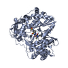
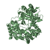
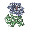
| Title | Crystal Structure of an Acetylacetone Dioxygenase from Acinetobacter johnsonii | ||||||
 Components Components | Acetylacetone-cleaving enzyme | ||||||
 Keywords Keywords | OXIDOREDUCTASE / JELLY ROLL / TETRAMER / Dioxygenase / Iron / Metal-binding | ||||||
| Function / homology |  Function and homology information Function and homology informationacetylacetone-cleaving enzyme / acetylacetone-cleaving enzyme activity / metal ion binding Similarity search - Function | ||||||
| Biological species |  Acinetobacter johnsonii (bacteria) Acinetobacter johnsonii (bacteria) | ||||||
| Method |  X-RAY DIFFRACTION / SYNCHROTRON / MAD / Resolution: 1.95 Å X-RAY DIFFRACTION / SYNCHROTRON / MAD / Resolution: 1.95 Å | ||||||
 Authors Authors | Stranzl, G.R. / Wagner, U.G. / Straganz, G. / Steiner, W. / Kratky, C. | ||||||
 Citation Citation | Journal: To be Published Title: Crystal Structure of an Acetylacetone Dioxygenase from Acinetobacter johnsonii Authors: Stranzl, G.R. / Wagner, U.G. / Straganz, G. / Steiner, W. / Kratky, C. | ||||||
| History |
|
- Structure visualization
| Structure viewer | Molecule: MolmilJmol/JSmol |
|---|
- Downloads & links
-Download
| PDBx/mmCIF format | 3bal.cif.gz | 133.6 KB | Display | PDBx/mmCIF format |
|---|---|---|---|---|
| PDB format | pdb3bal.ent.gz | 104.2 KB | Display | PDB format |
| PDBx/mmJSON format | 3bal.json.gz | Tree view | PDBx/mmJSON format | |
| Others |  Other downloads Other downloads |
-Validation report
| Arichive directory | https://data.pdbj.org/pub/pdb/validation_reports/ba/3balftp://data.pdbj.org/pub/pdb/validation_reports/ba/3bal | HTTPS FTP |
|---|
-Related structure data
| Similar structure data |
|---|


-Links
 PDBj
PDBj- Assembly
| Deposited unit | 
| ||||||||
|---|---|---|---|---|---|---|---|---|---|
| 1 |
| ||||||||
| Unit cell |
|
-Components
| #1: Protein | Mass: 16621.279 Da / Num. of mol.: 4 / Fragment: ACETYLACETONE DIOXYGENASE / Source method: isolated from a natural source / Source: (natural) Acinetobacter johnsonii (bacteria) / References: UniProt: Q8GNT2, acetylacetone-cleaving enzyme#2: Chemical | ChemComp-ZN /   Mass: 65.409 Da / Num. of mol.: 4 / Source method: obtained synthetically / Formula: Zn Mass: 65.409 Da / Num. of mol.: 4 / Source method: obtained synthetically / Formula: Zn#3: Water | ChemComp-HOH / |  Mass: 18.015 Da / Num. of mol.: 461 / Source method: isolated from a natural source / Formula: H2O Mass: 18.015 Da / Num. of mol.: 461 / Source method: isolated from a natural source / Formula: H2O |
|---|
-Experimental details
-Experiment
| Experiment | Method: X-RAY DIFFRACTION / Number of used crystals: 2 |
|---|
- Sample preparation
| Crystal | Density Matthews: 2.03 Å3/Da / Density % sol: 39.38 % |
|---|---|
| Crystal grow | Temperature: 277 K / Method: vapor diffusion, hanging drop / pH: 8 Details: 0.1M Tris HCl, 40% PEG 3350, 0.2M Sodium acetate, pH 8.0, VAPOR DIFFUSION, HANGING DROP, temperature 277K |
-Data collection
| Diffraction |
| ||||||||||||||||||||||||||||||||||||||||||||||||||||||||||||||||||||||||||||||||||||||||||||||||
|---|---|---|---|---|---|---|---|---|---|---|---|---|---|---|---|---|---|---|---|---|---|---|---|---|---|---|---|---|---|---|---|---|---|---|---|---|---|---|---|---|---|---|---|---|---|---|---|---|---|---|---|---|---|---|---|---|---|---|---|---|---|---|---|---|---|---|---|---|---|---|---|---|---|---|---|---|---|---|---|---|---|---|---|---|---|---|---|---|---|---|---|---|---|---|---|---|---|
| Diffraction source |
| ||||||||||||||||||||||||||||||||||||||||||||||||||||||||||||||||||||||||||||||||||||||||||||||||
| Detector |
| ||||||||||||||||||||||||||||||||||||||||||||||||||||||||||||||||||||||||||||||||||||||||||||||||
| Radiation |
| ||||||||||||||||||||||||||||||||||||||||||||||||||||||||||||||||||||||||||||||||||||||||||||||||
| Radiation wavelength |
| ||||||||||||||||||||||||||||||||||||||||||||||||||||||||||||||||||||||||||||||||||||||||||||||||
| Reflection | Av σ(I) over netI: 13.1 / Number: 104251 / Rmerge(I) obs: 0.053 / Χ2: 1.15 / D res high: 1.95 Å / D res low: 25 Å / Num. obs: 36543 / % possible obs: 96.4 | ||||||||||||||||||||||||||||||||||||||||||||||||||||||||||||||||||||||||||||||||||||||||||||||||
| Diffraction reflection shell |
| ||||||||||||||||||||||||||||||||||||||||||||||||||||||||||||||||||||||||||||||||||||||||||||||||
| Reflection | Resolution: 1.95→25 Å / Num. obs: 37241 / % possible obs: 97.8 % / Rmerge(I) obs: 0.036 / Χ2: 0.96 / Net I/σ(I): 16.1 | ||||||||||||||||||||||||||||||||||||||||||||||||||||||||||||||||||||||||||||||||||||||||||||||||
| Reflection shell | Resolution: 1.95→2 Å / Rmerge(I) obs: 0.113 / Num. unique all: 2245 / Χ2: 0.613 / % possible all: 88.2 |

-Phasing
| Phasing | Method: MAD | |||||||||||||||||||||||||||||||||||
|---|---|---|---|---|---|---|---|---|---|---|---|---|---|---|---|---|---|---|---|---|---|---|---|---|---|---|---|---|---|---|---|---|---|---|---|---|
| Phasing set |
| |||||||||||||||||||||||||||||||||||
| Phasing MAD | D res high: 1.9 Å / D res low: 25 Å / FOM : 0.39 / Reflection: 36273 | |||||||||||||||||||||||||||||||||||
| Phasing MAD set |
| |||||||||||||||||||||||||||||||||||
| Phasing MAD set site |
| |||||||||||||||||||||||||||||||||||
| Phasing MAD shell |
| |||||||||||||||||||||||||||||||||||
| Phasing dm | FOM : 0 / FOM acentric: 0 / FOM centric: 0 / Reflection acentric: 0 / Reflection centric: 0 | |||||||||||||||||||||||||||||||||||
| Phasing dm shell |
|
- Processing
| Software |
| ||||||||||||||||||||||||||||||||||||||||
|---|---|---|---|---|---|---|---|---|---|---|---|---|---|---|---|---|---|---|---|---|---|---|---|---|---|---|---|---|---|---|---|---|---|---|---|---|---|---|---|---|---|
| Refinement | Method to determine structure: MAD / Resolution: 1.95→25 Å / FOM work R set: 0.9 / Cross valid method: THROUGHOUT / σ(F): 0 / Stereochemistry target values: Engh & Huber
| ||||||||||||||||||||||||||||||||||||||||
| Solvent computation | Bsol: 49.042 Å2 | ||||||||||||||||||||||||||||||||||||||||
| Displacement parameters | Biso mean: 20.486 Å2
| ||||||||||||||||||||||||||||||||||||||||
| Refinement step | Cycle: LAST / Resolution: 1.95→25 Å
| ||||||||||||||||||||||||||||||||||||||||
| Refine LS restraints |
| ||||||||||||||||||||||||||||||||||||||||
| Xplor file |
|
