 Movie
Movie Controller

[English] 日本語
 Yorodumi
- PDB-6sqx: Insights into a novel NlpC/P60 Endopeptidase from Photobacterium ... -
+ Open data

- Basic information
| Entry | Database: PDB / ID: 6sqx | ||||||
|---|---|---|---|---|---|---|---|
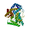
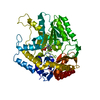
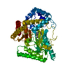
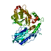
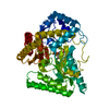
| Title | Insights into a novel NlpC/P60 Endopeptidase from Photobacterium damselae subsp. piscicida | ||||||
 Components Components | Peptide-binding protein | ||||||
 Keywords Keywords | HYDROLASE / NlpC/P60 / cell wall hydrolases / Crystallography X-Ray / D / L-endopeptidase | ||||||
| Function / homology |  Function and homology information Function and homology informationUncharacterised peptidase with NlpC/P60 domain, YkfC type / SH3b1 domain / NLPC_P60 stabilising domain, N term / SH3 domain (SH3b1 type) / : / NlpC/P60 family / NlpC/P60 domain profile. / Endopeptidase, NLPC/P60 domain / Papain-like cysteine peptidase superfamily Similarity search - Domain/homology | ||||||
| Biological species |  Photobacterium damsela subsp. piscicida (bacteria) Photobacterium damsela subsp. piscicida (bacteria) | ||||||
| Method |  X-RAY DIFFRACTION / SYNCHROTRON / MOLECULAR REPLACEMENT / Resolution: 1.4 Å X-RAY DIFFRACTION / SYNCHROTRON / MOLECULAR REPLACEMENT / Resolution: 1.4 Å | ||||||
 Authors Authors | Lisboa, J. / Pereira, P.J.B. / dos Santos, N.M.S. | ||||||
| Funding support |  Portugal, 1items Portugal, 1items
| ||||||
 Citation Citation | Journal: Msphere / Year: 2021 Title: A Secreted NlpC/P60 Endopeptidase from Photobacterium damselae subsp. piscicida Cleaves the Peptidoglycan of Potentially Competing Bacteria. Authors: Lisboa, J. / Pereira, C. / Rifflet, A. / Ayala, J. / Terceti, M.S. / Barca, A.V. / Rodrigues, I. / Pereira, P.J.B. / Osorio, C.R. / Garcia-Del Portillo, F. / Gomperts Boneca, I. / do Vale, A. ...Authors: Lisboa, J. / Pereira, C. / Rifflet, A. / Ayala, J. / Terceti, M.S. / Barca, A.V. / Rodrigues, I. / Pereira, P.J.B. / Osorio, C.R. / Garcia-Del Portillo, F. / Gomperts Boneca, I. / do Vale, A. / Dos Santos, N.M.S. | ||||||
| History |
|
- Structure visualization
| Structure viewer | Molecule: MolmilJmol/JSmol |
|---|
- Downloads & links
-Download
| PDBx/mmCIF format | 6sqx.cif.gz | 418.8 KB | Display | PDBx/mmCIF format |
|---|---|---|---|---|
| PDB format | pdb6sqx.ent.gz | 340.5 KB | Display | PDB format |
| PDBx/mmJSON format | 6sqx.json.gz | Tree view | PDBx/mmJSON format | |
| Others |  Other downloads Other downloads |
-Validation report
| Arichive directory | https://data.pdbj.org/pub/pdb/validation_reports/sq/6sqxftp://data.pdbj.org/pub/pdb/validation_reports/sq/6sqx | HTTPS FTP |
|---|
-Related structure data
| Related structure data |  3m1uS S: Starting model for refinement |
|---|---|
| Similar structure data |


-Links
 PDBj
PDBj


- Assembly
| Deposited unit | 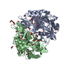
| ||||||||||
|---|---|---|---|---|---|---|---|---|---|---|---|
| 1 | 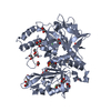
| ||||||||||
| 2 | 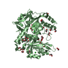
| ||||||||||
| Unit cell |
|
-Components
-Protein , 1 types, 2 molecules AB
| #1: Protein | Mass: 57393.453 Da / Num. of mol.: 2 Source method: isolated from a genetically manipulated source Details: Signal Peptide: M1-A19 LEHHHHHH: His Tag Source: (gene. exp.) Photobacterium damsela subsp. piscicida (bacteria)Gene: E4T26_07800, FA893_15790, PDPJ_2_00460 / Cell line (production host): Codon + / Production host: |
|---|
-Non-polymers , 5 types, 1100 molecules 


| #2: Chemical | ChemComp-CL /  Mass: 35.453 Da / Num. of mol.: 6 / Source method: obtained synthetically / Formula: Cl Mass: 35.453 Da / Num. of mol.: 6 / Source method: obtained synthetically / Formula: Cl#3: Chemical | ChemComp-EDO /  Mass: 62.068 Da / Num. of mol.: 38 / Source method: obtained synthetically / Formula: C2H6O2 Mass: 62.068 Da / Num. of mol.: 38 / Source method: obtained synthetically / Formula: C2H6O2#4: Chemical | ChemComp-PEG / |  Mass: 106.120 Da / Num. of mol.: 1 / Source method: obtained synthetically / Formula: C4H10O3 Mass: 106.120 Da / Num. of mol.: 1 / Source method: obtained synthetically / Formula: C4H10O3#5: Chemical | ChemComp-BCT / |  Mass: 61.017 Da / Num. of mol.: 1 / Source method: isolated from a natural source / Formula: CHO3 / Comment: pH buffer*YM Mass: 61.017 Da / Num. of mol.: 1 / Source method: isolated from a natural source / Formula: CHO3 / Comment: pH buffer*YM#6: Water | ChemComp-HOH / | Mass: 18.015 Da / Num. of mol.: 1054 / Source method: isolated from a natural source / Formula: H2O |
|---|
-Details
| Has ligand of interest | N |
|---|
-Experimental details
-Experiment
| Experiment | Method: X-RAY DIFFRACTION / Number of used crystals: 1 |
|---|
- Sample preparation
| Crystal | Density Matthews: 2.51 Å3/Da / Density % sol: 50.94 % |
|---|---|
| Crystal grow | Temperature: 293 K / Method: vapor diffusion, sitting drop / pH: 8 / Details: 100 mM Imidazole pH 8.0 + 15 % PEG 8K |
-Data collection
| Diffraction | Mean temperature: 100 K / Serial crystal experiment: N |
|---|---|
| Diffraction source | Source: SYNCHROTRON / Site: SOLEIL  / Beamline: PROXIMA 1 / Wavelength: 0.9786 Å / Beamline: PROXIMA 1 / Wavelength: 0.9786 Å |
| Detector | Type: DECTRIS PILATUS 6M / Detector: PIXEL / Date: Feb 12, 2016 |
| Radiation | Monochromator: Silicon Si crystal (111) / Protocol: SINGLE WAVELENGTH / Monochromatic (M) / Laue (L): M / Scattering type: x-ray |
| Radiation wavelength | Wavelength: 0.9786 Å / Relative weight: 1 |
| Reflection | Resolution: 1.4→38.3 Å / Num. obs: 216685 / % possible obs: 99.85 % / Redundancy: 5.5 % / CC1/2: 1 / Rmerge(I) obs: 0.046 / Rpim(I) all: 0.022 / Rrim(I) all: 0.051 / Net I/σ(I): 18.86 |
| Reflection shell | Resolution: 1.4→1.45 Å / Redundancy: 5.4 % / Rmerge(I) obs: 1.47 / Num. unique obs: 21426 / CC1/2: 0.616 / Rpim(I) all: 0.69 / Rrim(I) all: 1.626 / % possible all: 99.6 |
- Processing
| Software |
| |||||||||||||||||||||||||||||||||||||||||||||||||||||||||||||||||||||||||||||||||||||||||||||||||||||||||||||||||||||||||||||||||||||||||||||||||||||||||||||||||||||||||||||||||||||||||||||||||||||||||||||||||||||||||
|---|---|---|---|---|---|---|---|---|---|---|---|---|---|---|---|---|---|---|---|---|---|---|---|---|---|---|---|---|---|---|---|---|---|---|---|---|---|---|---|---|---|---|---|---|---|---|---|---|---|---|---|---|---|---|---|---|---|---|---|---|---|---|---|---|---|---|---|---|---|---|---|---|---|---|---|---|---|---|---|---|---|---|---|---|---|---|---|---|---|---|---|---|---|---|---|---|---|---|---|---|---|---|---|---|---|---|---|---|---|---|---|---|---|---|---|---|---|---|---|---|---|---|---|---|---|---|---|---|---|---|---|---|---|---|---|---|---|---|---|---|---|---|---|---|---|---|---|---|---|---|---|---|---|---|---|---|---|---|---|---|---|---|---|---|---|---|---|---|---|---|---|---|---|---|---|---|---|---|---|---|---|---|---|---|---|---|---|---|---|---|---|---|---|---|---|---|---|---|---|---|---|---|---|---|---|---|---|---|---|---|---|---|---|---|---|---|---|---|
| Refinement | Method to determine structure: MOLECULAR REPLACEMENT Starting model: 3m1u Resolution: 1.4→38.29 Å / SU ML: 0.17 / Cross valid method: THROUGHOUT / σ(F): 1.33 / Phase error: 19.61
| |||||||||||||||||||||||||||||||||||||||||||||||||||||||||||||||||||||||||||||||||||||||||||||||||||||||||||||||||||||||||||||||||||||||||||||||||||||||||||||||||||||||||||||||||||||||||||||||||||||||||||||||||||||||||
| Solvent computation | Shrinkage radii: 0.9 Å / VDW probe radii: 1.11 Å | |||||||||||||||||||||||||||||||||||||||||||||||||||||||||||||||||||||||||||||||||||||||||||||||||||||||||||||||||||||||||||||||||||||||||||||||||||||||||||||||||||||||||||||||||||||||||||||||||||||||||||||||||||||||||
| Displacement parameters | Biso max: 105.92 Å2 / Biso mean: 28.36 Å2 / Biso min: 12.61 Å2 | |||||||||||||||||||||||||||||||||||||||||||||||||||||||||||||||||||||||||||||||||||||||||||||||||||||||||||||||||||||||||||||||||||||||||||||||||||||||||||||||||||||||||||||||||||||||||||||||||||||||||||||||||||||||||
| Refinement step | Cycle: final / Resolution: 1.4→38.29 Å
| |||||||||||||||||||||||||||||||||||||||||||||||||||||||||||||||||||||||||||||||||||||||||||||||||||||||||||||||||||||||||||||||||||||||||||||||||||||||||||||||||||||||||||||||||||||||||||||||||||||||||||||||||||||||||
| LS refinement shell | Refine-ID: X-RAY DIFFRACTION / Rfactor Rfree error: 0 / Total num. of bins used: 30
|
