 Movie
Movie Controller

[English] 日本語
 Yorodumi
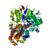
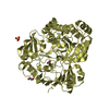
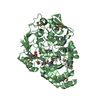
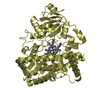
- PDB-4zei: PBP AccA from A. tumefaciens C58 in complex with L-arabinose-2-ph... -
+ Open data

- Basic information
| Entry | Database: PDB / ID: 4zei | ||||||
|---|---|---|---|---|---|---|---|
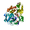
| Title | PBP AccA from A. tumefaciens C58 in complex with L-arabinose-2-phosphate | ||||||
 Components Components | ABC transporter, substrate binding protein (Agrocinopines A and B) | ||||||
 Keywords Keywords | TRANSPORT PROTEIN / PBP / CLASS C | ||||||
| Function / homology |  Function and homology information Function and homology informationpeptide transport / peptide transmembrane transporter activity / ATP-binding cassette (ABC) transporter complex / outer membrane-bounded periplasmic space Similarity search - Function | ||||||
| Biological species |  Agrobacterium tumefaciens (bacteria) Agrobacterium tumefaciens (bacteria) | ||||||
| Method |  X-RAY DIFFRACTION / SYNCHROTRON / MOLECULAR REPLACEMENT / Resolution: 2.3 Å X-RAY DIFFRACTION / SYNCHROTRON / MOLECULAR REPLACEMENT / Resolution: 2.3 Å | ||||||
 Authors Authors | El Sahili, A. / Morera, S. | ||||||
 Citation Citation | Journal: Plos Pathog. / Year: 2015 Title: A Pyranose-2-Phosphate Motif Is Responsible for Both Antibiotic Import and Quorum-Sensing Regulation in Agrobacterium tumefaciens. Authors: El Sahili, A. / Li, S.Z. / Lang, J. / Virus, C. / Planamente, S. / Ahmar, M. / Guimaraes, B.G. / Aumont-Nicaise, M. / Vigouroux, A. / Soulere, L. / Reader, J. / Queneau, Y. / Faure, D. / Morera, S. | ||||||
| History |
|
- Structure visualization
| Structure viewer | Molecule: MolmilJmol/JSmol |
|---|
- Downloads & links
-Download
| PDBx/mmCIF format | 4zei.cif.gz | 217.4 KB | Display | PDBx/mmCIF format |
|---|---|---|---|---|
| PDB format | pdb4zei.ent.gz | 172.5 KB | Display | PDB format |
| PDBx/mmJSON format | 4zei.json.gz | Tree view | PDBx/mmJSON format | |
| Others |  Other downloads Other downloads |
-Validation report
| Arichive directory | https://data.pdbj.org/pub/pdb/validation_reports/ze/4zeiftp://data.pdbj.org/pub/pdb/validation_reports/ze/4zei | HTTPS FTP |
|---|
-Related structure data
| Related structure data |  4ra1C 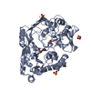 4ze8C  4ze9C  4zebC 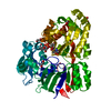 4zecC 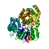 4zedC  4zekC  4oup C: citing same article ( S: Starting model for refinement |
|---|---|
| Similar structure data |


-Links
 PDBj
PDBj

- Assembly
| Deposited unit | 
| ||||||||
|---|---|---|---|---|---|---|---|---|---|
| 1 |
| ||||||||
| Unit cell |
|
-Components
| #1: Protein | Mass: 57178.102 Da / Num. of mol.: 1 Source method: isolated from a genetically manipulated source Source: (gene. exp.) Agrobacterium tumefaciens (bacteria) / Gene: accA, Atu6139 / Production host: | ||
|---|---|---|---|
| #2: Sugar | ChemComp-LAO / 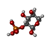  Type: L-saccharide, alpha linking / Mass: 230.110 Da / Num. of mol.: 1 Type: L-saccharide, alpha linking / Mass: 230.110 Da / Num. of mol.: 1Source method: isolated from a genetically manipulated source Formula: C5H11O8P | ||
| #3: Chemical | ChemComp-EDO /   Mass: 62.068 Da / Num. of mol.: 5 / Source method: obtained synthetically / Formula: C2H6O2 Mass: 62.068 Da / Num. of mol.: 5 / Source method: obtained synthetically / Formula: C2H6O2#4: Water | ChemComp-HOH / |  Mass: 18.015 Da / Num. of mol.: 144 / Source method: isolated from a natural source / Formula: H2O Mass: 18.015 Da / Num. of mol.: 144 / Source method: isolated from a natural source / Formula: H2O |
-Experimental details
-Experiment
| Experiment | Method: X-RAY DIFFRACTION / Number of used crystals: 1 |
|---|
- Sample preparation
| Crystal | Density Matthews: 2.15 Å3/Da / Density % sol: 42.81 % |
|---|---|
| Crystal grow | Temperature: 298 K / Method: vapor diffusion, hanging drop / pH: 5.6 Details: 20% PEG 4K, 0.2M ACNH4, 0.1M NA CITRATE PH 5.6, VAPOR DIFFUSION, HANGING DROP, TEMPERATURE 298K PH range: 5.6 |
-Data collection
| Diffraction | Mean temperature: 100 K |
|---|---|
| Diffraction source | Source: SYNCHROTRON / Site: SOLEIL  / Beamline: PROXIMA 1 / Wavelength: 0.987 Å / Beamline: PROXIMA 1 / Wavelength: 0.987 Å |
| Detector | Type: DECTRIS PILATUS 6M / Detector: PIXEL / Date: Dec 8, 2013 |
| Radiation | Monochromator: SI111 / Protocol: SINGLE WAVELENGTH / Monochromatic (M) / Laue (L): M / Scattering type: x-ray |
| Radiation wavelength | Wavelength: 0.987 Å / Relative weight: 1 |
| Reflection | Resolution: 2.3→50 Å / Num. obs: 22254 / % possible obs: 99.6 % / Observed criterion σ(I): 1 / Redundancy: 4.4 % / Biso Wilson estimate: 55.82 Å2 / Rsym value: 0.146 / Net I/σ(I): 8.48 |
| Reflection shell | Resolution: 2.3→2.44 Å / Rsym value: 1.17 / % possible all: 96.6 |
- Processing
| Software |
| ||||||||||||||||||||||||||||||||||||||||||||||||||||||||||||||||||||||||||||||||||||||||||||||||||||||||||||||||||
|---|---|---|---|---|---|---|---|---|---|---|---|---|---|---|---|---|---|---|---|---|---|---|---|---|---|---|---|---|---|---|---|---|---|---|---|---|---|---|---|---|---|---|---|---|---|---|---|---|---|---|---|---|---|---|---|---|---|---|---|---|---|---|---|---|---|---|---|---|---|---|---|---|---|---|---|---|---|---|---|---|---|---|---|---|---|---|---|---|---|---|---|---|---|---|---|---|---|---|---|---|---|---|---|---|---|---|---|---|---|---|---|---|---|---|---|
| Refinement | Method to determine structure: MOLECULAR REPLACEMENT Starting model: 4OUP 4oup Resolution: 2.3→25.09 Å / Cor.coef. Fo:Fc: 0.94 / Cor.coef. Fo:Fc free: 0.909 / SU R Cruickshank DPI: 0.391 / Cross valid method: THROUGHOUT / σ(F): 0
| ||||||||||||||||||||||||||||||||||||||||||||||||||||||||||||||||||||||||||||||||||||||||||||||||||||||||||||||||||
| Displacement parameters | Biso mean: 50.25 Å2
| ||||||||||||||||||||||||||||||||||||||||||||||||||||||||||||||||||||||||||||||||||||||||||||||||||||||||||||||||||
| Refine analyze | Luzzati coordinate error obs: 0.33 Å | ||||||||||||||||||||||||||||||||||||||||||||||||||||||||||||||||||||||||||||||||||||||||||||||||||||||||||||||||||
| Refinement step | Cycle: LAST / Resolution: 2.3→25.09 Å
| ||||||||||||||||||||||||||||||||||||||||||||||||||||||||||||||||||||||||||||||||||||||||||||||||||||||||||||||||||
| Refine LS restraints |
| ||||||||||||||||||||||||||||||||||||||||||||||||||||||||||||||||||||||||||||||||||||||||||||||||||||||||||||||||||
| LS refinement shell | Resolution: 2.3→2.41 Å / Total num. of bins used: 11
| ||||||||||||||||||||||||||||||||||||||||||||||||||||||||||||||||||||||||||||||||||||||||||||||||||||||||||||||||||
| Refinement TLS params. | Method: refined / Origin x: -16.9967 Å / Origin y: -26.6891 Å / Origin z: 14.1511 Å
| ||||||||||||||||||||||||||||||||||||||||||||||||||||||||||||||||||||||||||||||||||||||||||||||||||||||||||||||||||
| Refinement TLS group | Selection details: A 32 A 521 |
